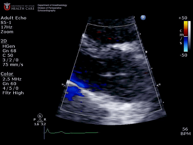
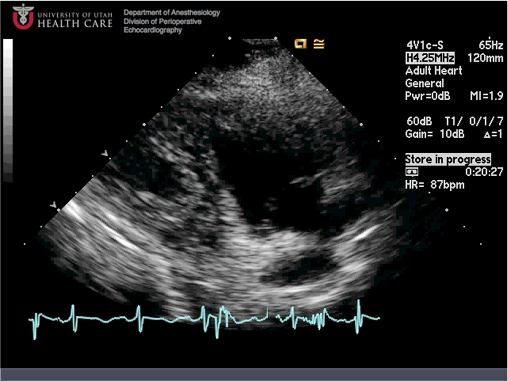
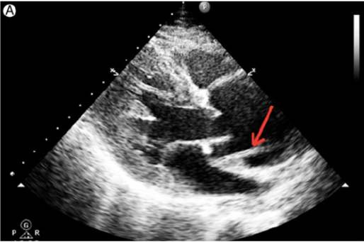
What are we looking at here?
![]()
- Aortic valve zoom from apical window, aortic regurgitation
- Mitral valve zoom from apical window, normal colour
- Aortic valve zoon from parasternal window, normal colour flow
- Pulmonic valve zoom from parasternal window, normal colour flow
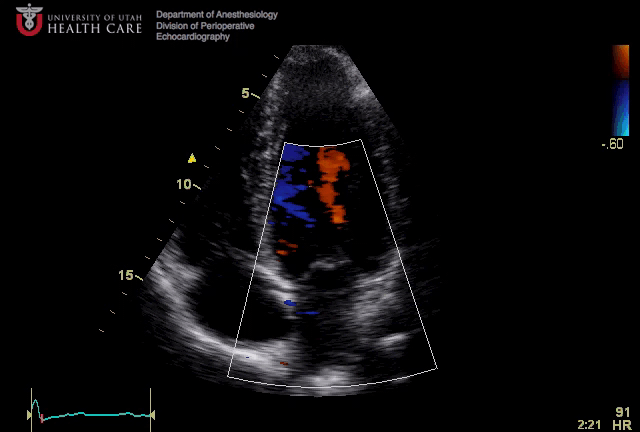
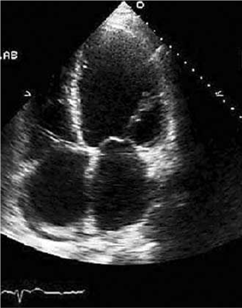
What view is this? Is the colour flow doppler set appropriately?
![]()
- Apical 4 chamber, appropriate colour flow Doppler settings on tricuspid valve
- Subcoastal 4 chamber view, inappropriate colour flow Doppler settings on mitral valve
- Apical 4 chamber, appropriate colour flow Doppler settings on mitral valve
- Apical 4 chamber, inappropriate colour flow Doppler settings on mitral valve
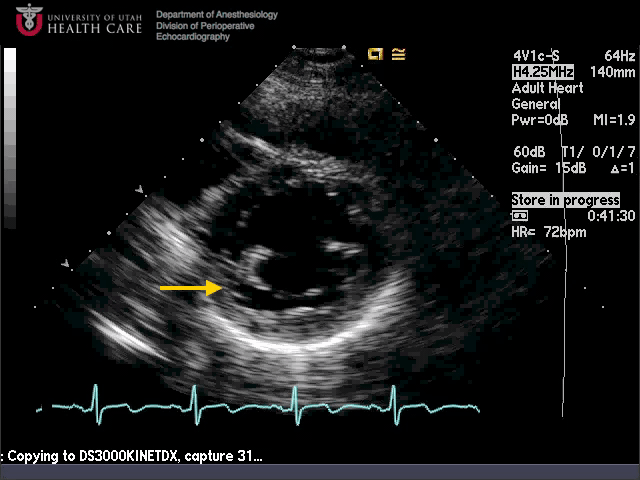
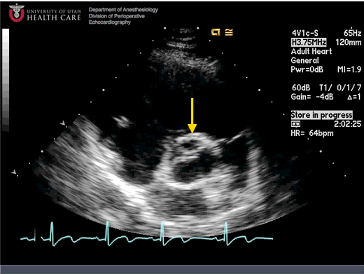
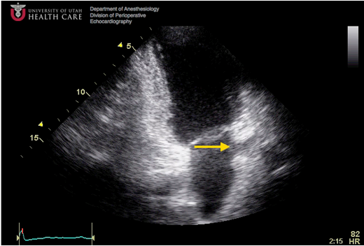
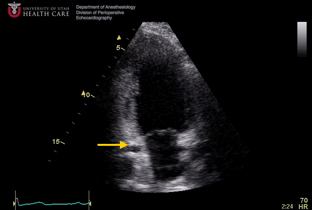
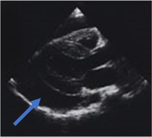
What view is this and what is the arrow pointing at?
![]()
- Parasternal LV short axis, basal inferior segment
- Subcostal LV short axis, basal inferior segment
- Parasternal LV short axis, basal anterior segment
- Subcostal LV short axis, mid inferolateral segment
- Parasternal LV short axis, mid anterior segment
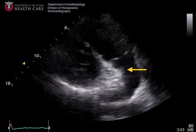
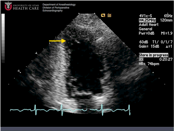
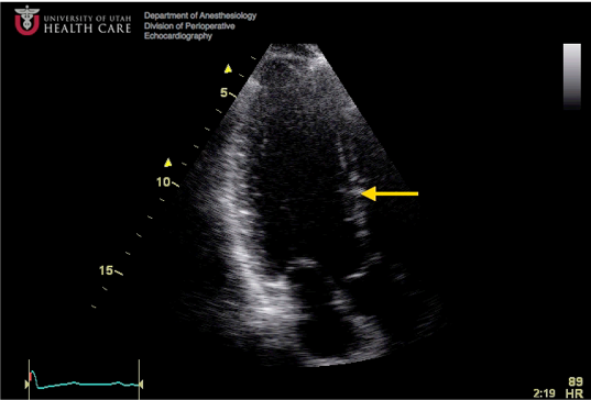
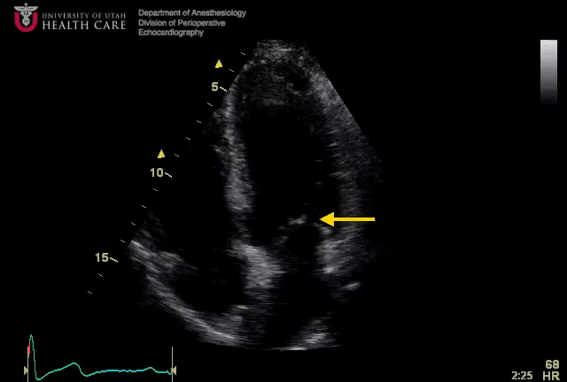
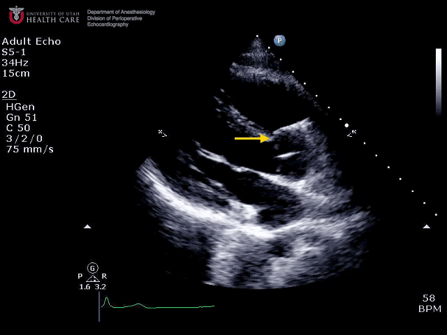
What view is this and what structure is shown by the arrow?
![]()
- Apical 4 chamber, left atrium
- Parasternal RV outflow, main PA
- Parasternal RV inflow, right atrium
- Apical 2 chamber, left atrium
What view is this and what is shown by the arrow?
![]()
- Apical RV inflow/outflow, right cusp of pulmonic valve
- Apical RV inflow/outflow, right coronary cusp of aortic valve
- Parasternal RV inflow/outflow, noncoronary cusp of aortic valve
- Apical RV inflow/outflow, left coronary cusp of aortic valve
- Parasternal RV inflow/outflow, right coronary cusp of aortic valve
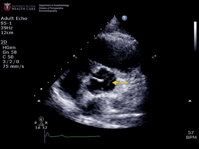
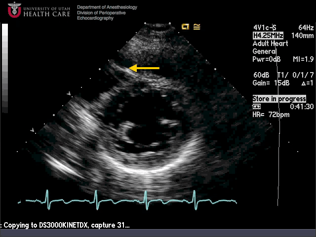
What view is this and what structure is shown by the arrow?
![]()
- Parasternal 2 chamber, apical inferolateral segment
- Apical 2 chamber, apical inferior segment
- Apical 2 chamber, apical anterior segment
- Parasternal 2 chamber, basal anteroseptal segment
Where is the arrow pointing?
![]()
- Left cusp of pulmonic valve
- Left coronary cusp of AV
- Non coronary cusp of AV
- Right coronary cusp of AV
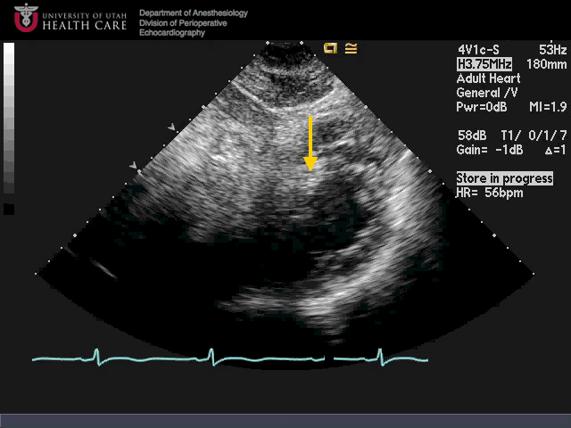
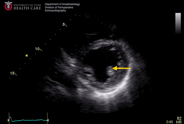
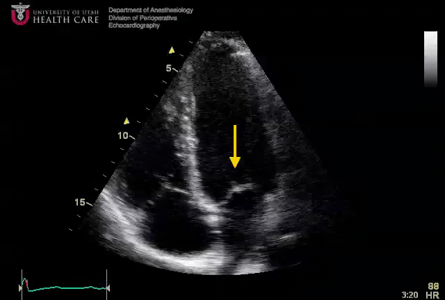
What view is this (careful) and what structure is shown with the arrow?
![]()
- Parasternal LV short axis, mid anteroseptal segment
- Parasternal LV short axis, mid inferoseptal segment
- Subcostal LV short axis, mid inferoseptal segment
- Transgastric LV short axis, apical septal segment
- Subcostal LV short axis, mid anteroseptal segment
What are we looking at here?
![]()
- Subcostal long axis, aortic valve
- Parasternal long axis, mid anteroseptal segment
- Parasternal long axis, mid inferolateral segment
- Apical long axis, basal anteroseptal segment
- Apical long axis, basal inferoseptal segment
What view is this and what is the arrow pointing at?
![]()
- Transgastric LV short axis, posteromedial papillary muscle
- Parasternal LV short axis, posteromedial papillary muscle
- Parasternal LV short axis, anterolateral papillary muscle
- Transgastric LV short axis, anterolateral papillary muscle
What view is this and what structure is shown by the arrow?
![]()
- Parasternal LV short axis, midpapillary level, mid anteroseptal segment
- Subcostal LV short axis, midpapillary level, midinferoseptal segment
- Parasternal LV short axis, apical level, apical septal segment
- Subcostal LV short axis, apical level, apical septal segment
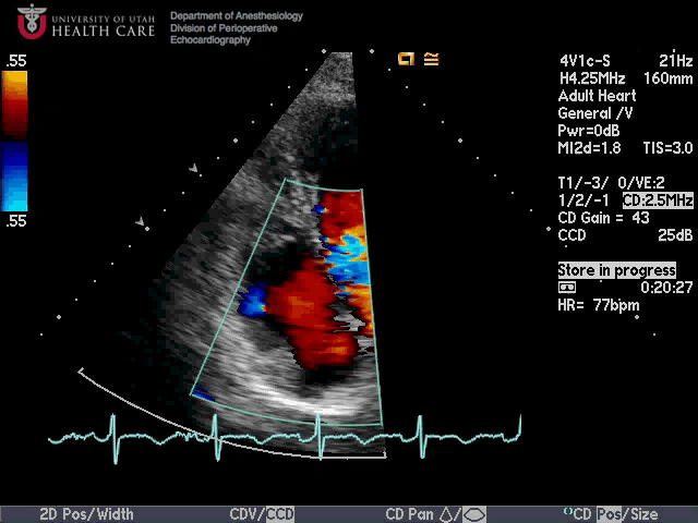
What view is this and what is seen?
![]()
- Parasternal long axis, color flow across the aortic valve concerning for stenosis
- Apical long axis, color flow across the aortic valve concerning for dynamic outflow obstruction
- Parasternal long axis, normal color flow across the aortic valve
- Parasternal long axis, color flow across the aortic valve concerning for insufficiency
What view is this? Describe the global right ventricular systolic function.
![]()
- Parasternal 4 chamber, reduced RV function
- Apical 4 chamber, reduced RV function
- Apical 4 chamber, normal RV function
- Subcostal 4 chamber, normal RV function
- Subcostal 4 chamber, reduced RV function
What view is this and what structure is shown by the arrow?
![]()
- Apical 4 chamber, left lower pulmonary vein
- Apical 2 chamber, left atrial appendage
- Apical 2 chamber, coronary sinus
- Apical long axis, thoracic aorta
- Apical long axis, aortic valve
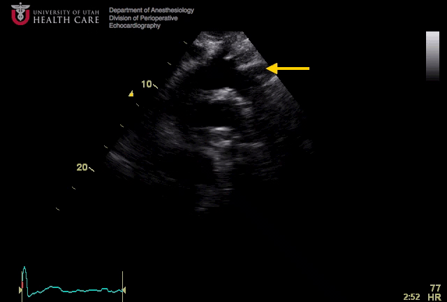
What view is this and what is shown by the arrow?
![]()
- Suprasternal aortic arch long axis, innominate artery
- Parasternal aortic long axis, right common carotid artery
- Suprasternal aortic arch long axis, left subclavian artery
- Parasternal aortic long axis, right subclavian artery
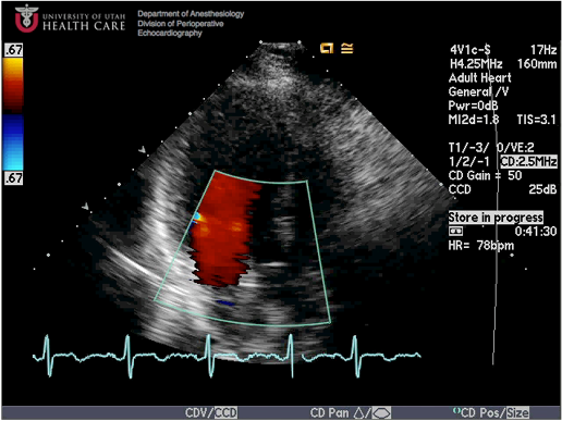
What view is this and what do we see?
![]()
- Parasternal long axis, normal mitral valve color flow
- Parasternal RV inflow, normal tricuspid valve color flow
- Parasternal long axis, mitral valve color flow with significant regurgitation
- Parasternal RV inflow, significant tricuspid regurgitation
What view is this and what are we seeing?
![]()
- Apical long axis, normal colour flow
- Apical long axis, significant mitral regurgitation
- Parasternal long axis, significant aortic insufficiency
- Parasternal long axis, normal colour flow
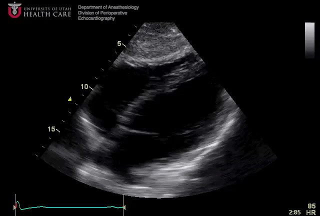
What view is this?
![]()
- Parasternal RV outflow
- Parasternal RV inflow
- Apical long axis
- Subcostal 4 chamber
- Transgastric RV inflow-outflow
What view is this? Where are all the Zs?
![]()
- Apical LV short axis, basal inferoseptal and basal anterior segments
- Subcostal LV short axis, apical inferior and apical anterior segments
- Parasternal LV short axis, basal inferoseptal and basal anterolateral segments
- Parasternal LV short axis, mid inferoseptal and mid anterolateral segments
What view is this and what structure is shown by the arrow?
![]()
- Apical 4 chamber, right atrium
- Parasternal long axis, aorta
- Apical long axis, right atrium
- Apical 2 chamber, left atrial appendage
- Apical 2 chamber, coronary sinus
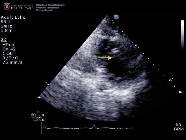
What view is this and what structure is indicated?
![]()
- Apical long axis, anterior mitral leaflet
- Apical 4 chamber, septal leaflet of tricuspid valve
- Apical long axis, aortic valve
- Apical 4 chamber, posterior mitral leaflet
- Apical 4 chamber, anterior mitral leaflet
What view is this?
![]()
- Parasternal long axis, normal mitral colour flow
- Subcostal 4 chamber, normal MV colour flow
- Parasternal RV inflow, normal tricuspid colour flow
- Apical 4 chamber, normal MV colour flow
- Apical 4 chamber, normal TV colour flow
What view is this and what is the arrow indicating?
![]()
- Parasternal long axis, right coronary cusp of the aortic valve
- Midesophageal long axis, left coronary cusp of the aortic valve
- Apical long axis, left coronary cusp of the aortic valve
- Midesophageal long axis, non-coronary cusp of the aortic valve
- Apical long axis, non-coronary cusp of the aortic valve
- Parasternal long axis, left coronary cusp of the aortic valve
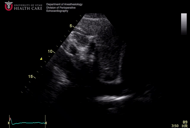
What view is this? What does this tell us about the patient’s hemodynamic state?
![]()
- Subcostal IVC long axis, low-normal right atrial pressure
- Subcostal IVC long axis, elevated right atrial pressure
- Subcostal aortic long axis, severe aortic insufficiency
- Subcostal aortic long axis, cardiac tamponade
What view is this? Is the RV function normal, and how large is the effusion?
![]()
- Subcostal 4 chamber, normal RV function, no effusion seen
- Parasternal long axis, reduced RV function, small pericardial effusion
- Apical 4 chamber, reduced RV function, moderate pericardial effusion
- Apical 4 chamber, normal RV function, no pericardial effusion
- Subcostal 4 chamber, reduced RV function, small pericardial effusion
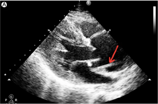
What view is this and what structure is indicated by the arrow?
![]()
- Subcostal 4 chamber, posterior mitral leaflet
- Subcostal 4 chamber, anterior mitral leaflet
- Apical 4 chamber, anterior mitral leaflet
- Apical 3 chamber, aortic valve
- Apical 4 chamber, posterior mitral leaflet
What view is this and what structure is shown by the arrow?
![]()
- Apical LV short axis, RV free wall
- Subcostal 4 chamber, RV free wall
- Subcostal 4 chamber, pericardial effusion
- Parasternal LV short axis, pulmonary embolus
- Parasternal LV short axis, moderator band
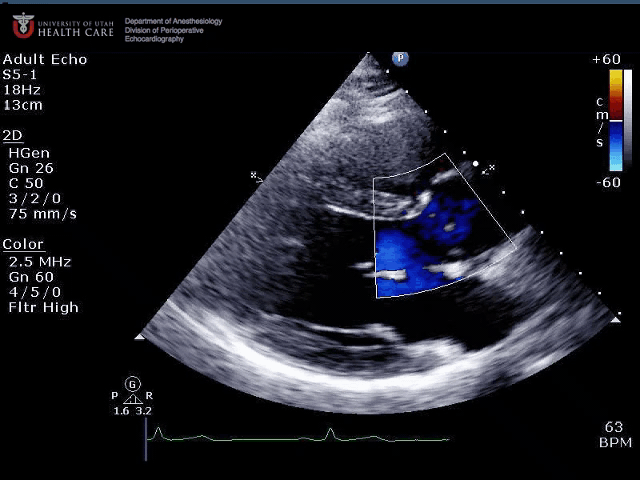
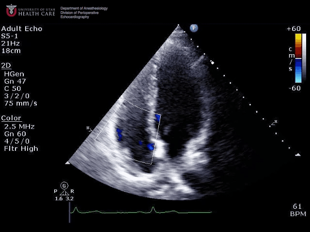
What view is this? Describe the colour flow Doppler findings.
![]()
- Apical 4 chamber, severe TR
- Subcostal 4 chamber, moderate TR
- Apical 4 chamber, mild TR
- Subcostal 4 chamber, mild TR
What view is this and what is shown by the arrow?
![]()
Parasternal long axis, Dissection flap
This patient is likely to have?
![]()
- Severe aortic stenosis (AS)
- Severe mitral regurgitation (MR)
- Severe pulmonary hypertension
- Mild AS
Risk of aortic dissection is increased in the following conditions except:
![]()
- Marfan’s syndrome
- Bicuspid aortic valve
- Pregnancy
- Mitral stenosis
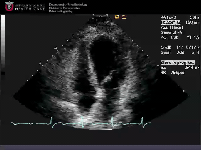
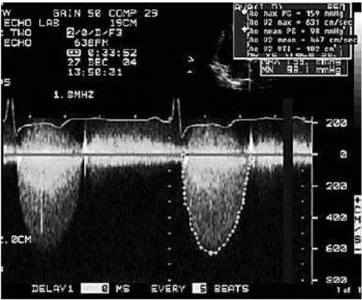
This is an end systolic frame in a patient with shortness of breath. What is the most likely diagnosis?
![]()
- Ebstein’s anomaly
- Hypertrophic cardiomyopathy
- Atrial septal defect
- Dilated cardiomyopathy
What does the arrow show?
![]()
Pericardial Effusion 心包积液