# Questions
Which of the following statements are true?
- Frequency = wavelength/velocity 频率 = 波长 / 速度
- The frequency of transthoracic echo is 2-3MHz 经胸超声频率为 2~3 MHz
- Increasing the frequency results in better penetration of ultrasound waves 提高频率可提高超声波的穿透性
- The velocity of ultrasound in cardiac tissue is 1570m/s 超声在心肌组织中的速度为 1570 m/s
- Attenuation is dependant on the frequency of ultrasound 衰减取决于超声波的频率
频率 = 速度 / 波长
增加频率时分辨率是增加的,但是穿透力是下降的。
超声在心肌组织中的传播速度是 1540 m/s
Which of the following statements are true?
- Specular reflection is produced by reflectors which are larger than the wavelength 反射是由大于波长的反射器产生的
- Scattered reflection is produced by reflectors which are smaller than the wavelength 散射是由小于波长的反射器产生的
- M-mode has a low sampling rate M 模式的采样率很低
- Piezo-electric crystals are not used in modern ultrasound probes 压电晶体不用于现代超声波探针
- High intensity signals appear black on ultrasound 高强度信号在超声波上显示为黑色
M 模式其实有很高的采样率,所以它的时间分辨率是很高的。
Which of the following statements are true?
- Axial resolution is the ability to separate two adjacent structures which are perpendicular to the beam 轴向分辨率是分辨和超声束相垂直的两个点的能力
- Lateral resolution is the ability to separate structures parallel to the ultrasound beam 横向分辨率是分辨与超声束相平行的结构的能力
- Axial resolution is increased at higher frequencies 在较高频率下,轴向分辨率增加
- Temporal resolution is the ability to track moving objects over time 时间分辨率是随着时间推移跟踪移动对象的能力
- Temporal resolution increases with a lower frame rate 帧频越低,时间分辨率越高
轴向分辨率是分辨和超声束相平行的两个点的能力,超声表现为上下两点
横向分辨率是分辨和超声束相垂直的结构的能力,在超声上表现为左右两点
帧速率越高,时间分辨率越高,所以可以减少感兴趣扇区的面积深度、宽度,这样可以提高帧频,从而提高时间分辨率
Which of the following statements are true?
- Aliasing occurs because the system is try to image an event occurring faster than the rate of sampling 产生混叠的原因是系统尝试以比采样速率更快的速度对发生的事件进行成像
- Aliasing affects continuous wave doppler more than pulse wave doppler 混叠对连续波多谱勒的影响大于脉冲波多谱勒
- Doppler echocardiography only gives information on speed of movement 多普勒超声心动图仅提供有关运动速度的信息
- Doppler signal should be perpendicular to the flow of blood to yield maximum velocity 多普勒信号应垂直于血流以产生最大速度
- Higher doppler frequency is obtained if there is lower blood velocity 血流速度越低,多谱勒频率越高
混叠影响脉冲多普勒多于连续多普勒。
多普勒超声心动图不但提供速度的信息,还提供方向的信息。
在做连续多普勒时,尽量要平行于血流的方向,因为它有角度依赖问题,这样才可以取得尽量高的速度。
较低的血流速度,一般取得的是较低的多普勒频谱。
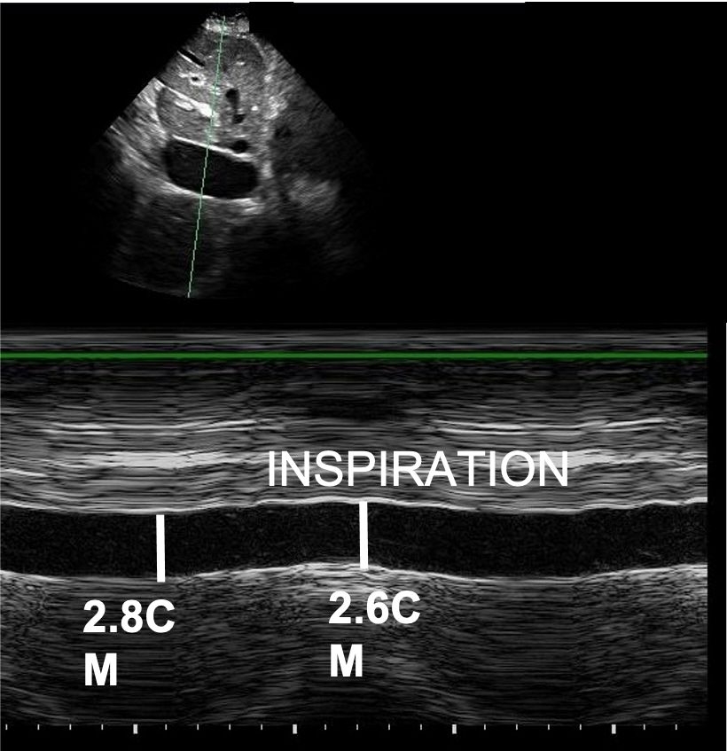
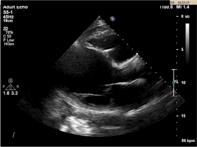
# Case 1
48 year old male
Background: HTN
Medications: Metformin
Presents with episode of collapse
Small right pleural effusion
ECG shows ST depression in inferolateral leads
Troponin is 120 (normal 0-40).
You are asked to perform an echocardiogram.


![]()
What is the right atrial pressure?
- 0-5mmHg
- 5-10mmHg
- 15-20mmHg
- 10-15mmHg
The Eccentricity index suggests:
- RV pressure overload
- RV volume overload
- Volume and pressure overload
- Neither
- Left ventricular impairment
What is the PASP?
- 46 – 51mmHg
- 36 – 41mmHg
- 30 – 35mmHg
- 25 – 30mmHg
- Unable to calculate based on the data
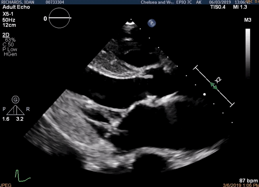
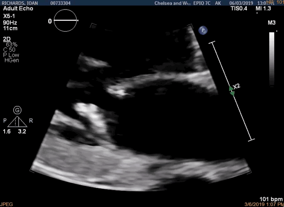
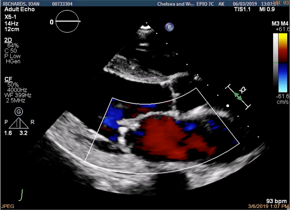
# Case 2
72F presents with breathlessness and peripheral oedema.
ECG: Atrial fibrillation
CXR: Pulmonary oedema.
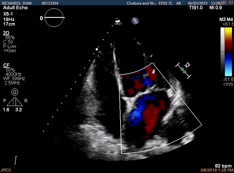
![]()
![]()
![]()
There is evidence of:
- Mild MR
- Moderate MR
- Severe MR
- Severe AR
What is the possible mechanism of the MR?
- Anterior leaflet prolapse
- Posterior leaflet prolapse
- Anterior leaflet endocarditis
- Posterior leaflet endocarditis
- Posterior leaflet flail
- Anterior leaflet flail
- Differential includes number 4 and 5
- Differential includes number 3 and 6
What is the MR?
![]()
- Anteriorly directed
- Posteriorly directed
- Central
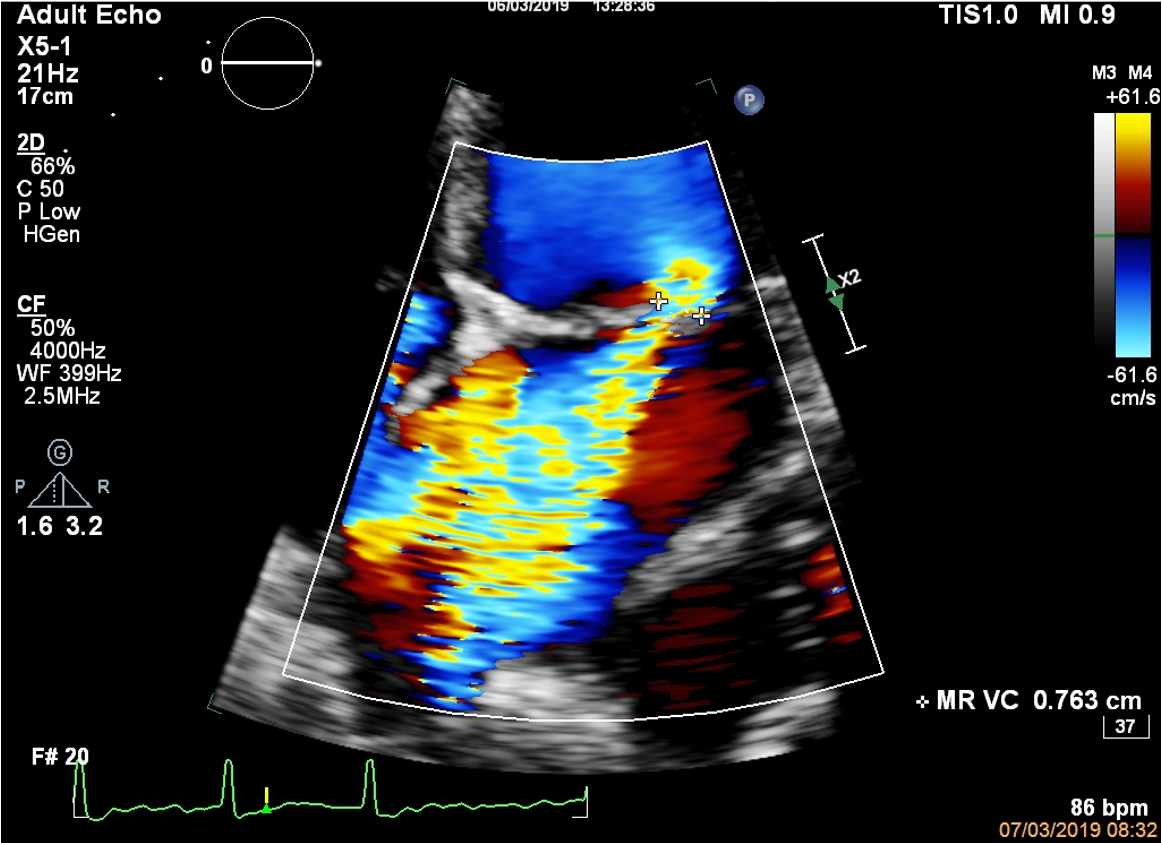
What does the vena contracta of 0.74 suggest?
![]()
- Mild MR
- Moderate MR
- Severe MR
- Not enough information
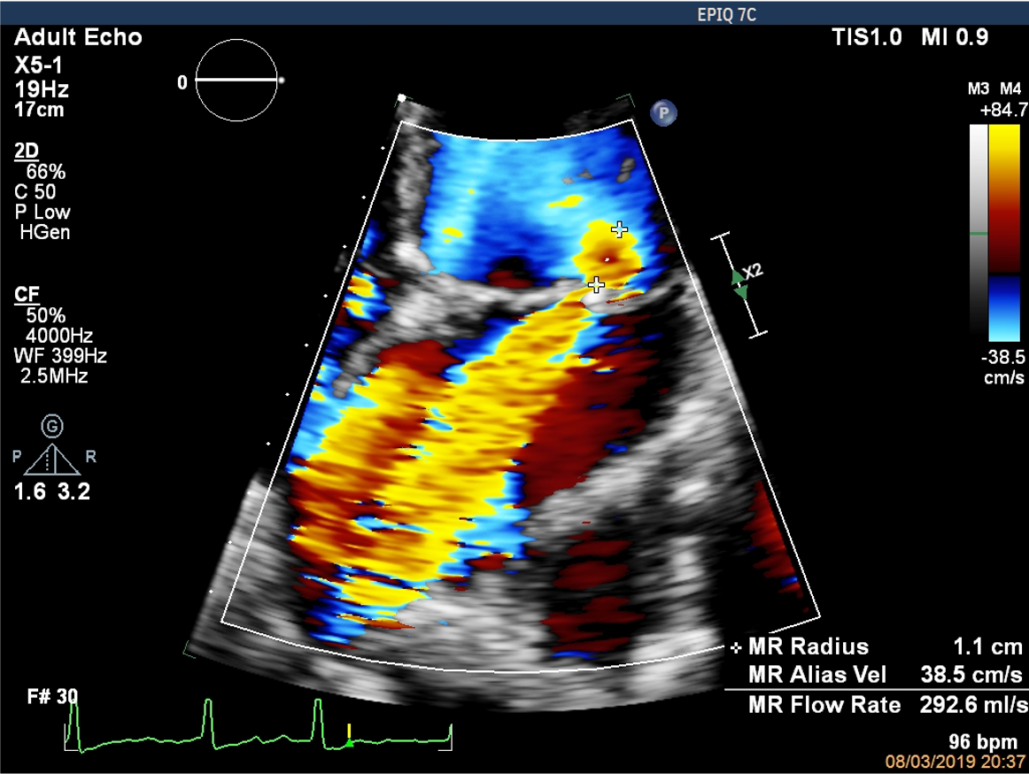
What does the PISA radius suggest?
![]()
- Mild MR
- Moderate MR
- Severe MR
- Not enough information
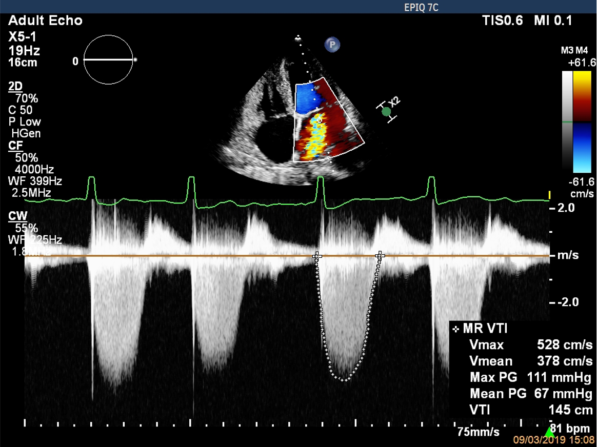
Based on the following data, what is the regurgitant volume?
![]()
What is the EROA?
- 0.55cm2
- 0.45cm2
- 0.35cm2
- 0.65cm2
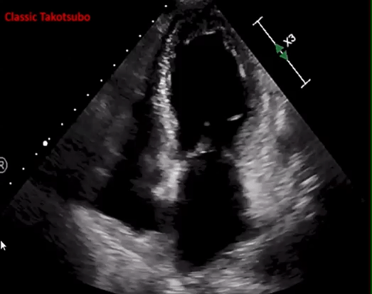
# Case 3
60 year old female
ECG: Anterior ST elevation
CXR: Mild pulmonary oedema.
![]()
What is this patient’s ejection fraction?
- EF 45-50%
- EF 40-45%
- EF 30-35%
- EF > 55%
- Diastolic dysfunction
The differential here includes the following:
- Restrictive CM
- Takotsubo CM
- Ischaemic CM
- Hypertrophic CM
- Normal
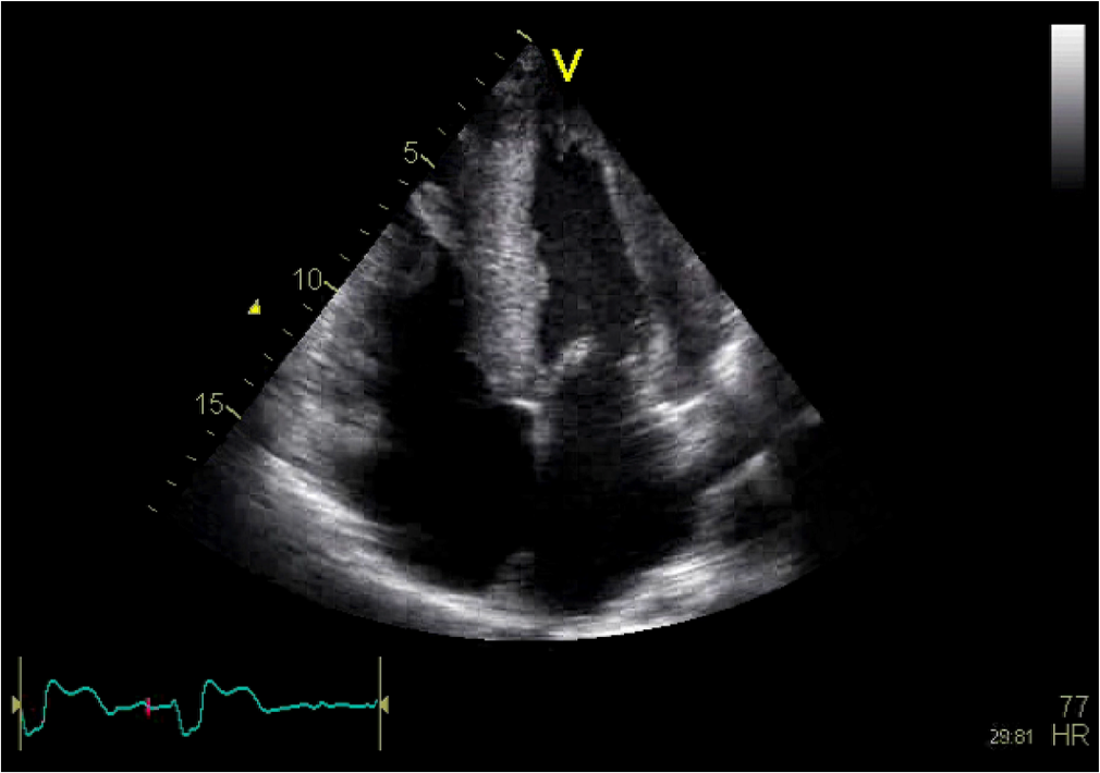
# Case 4
![]()
![]()
This patient has evidence of:
- Asymmetrical LVH
- Symmetrical LVH
- Dilated atria
- Normal longitudinal function
- Impaired ejection fraction
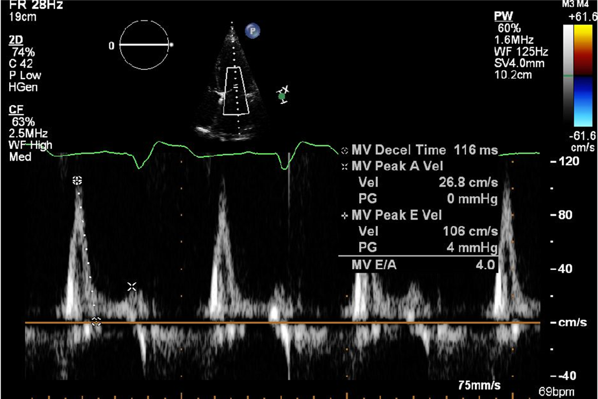
This MV inflow is suggestive of:
- Normal diastolic function
- Normal systolic function
- Mild systolic dysfunction
- Restrictive diastolic dysfunction
- Severe mitral regurgitation
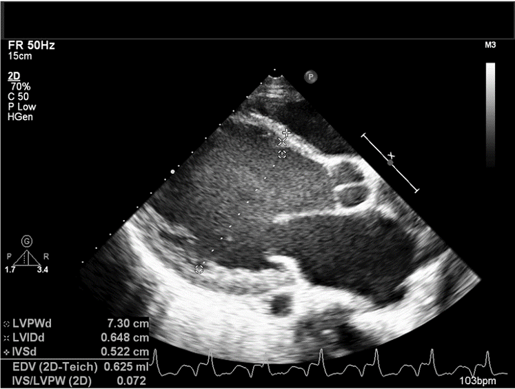
![]()
- The left ventricle is:
- Normal
- Sphericity index > 1.5
- Dilated
- Hypertrophied
![]()
- The LV function is:
- Normal
- 55 – 60%
- 40-45%
- 30-35%
- < 20%