# Case 1
73 year old female with chest pain
ECG: LVH
CXR: Unremarkable.
![]()
- What is the left ventricular function?
- > 55%
- 45 - 50%
- 35 - 40%
- 30 - 35%
- < 20%
![]()
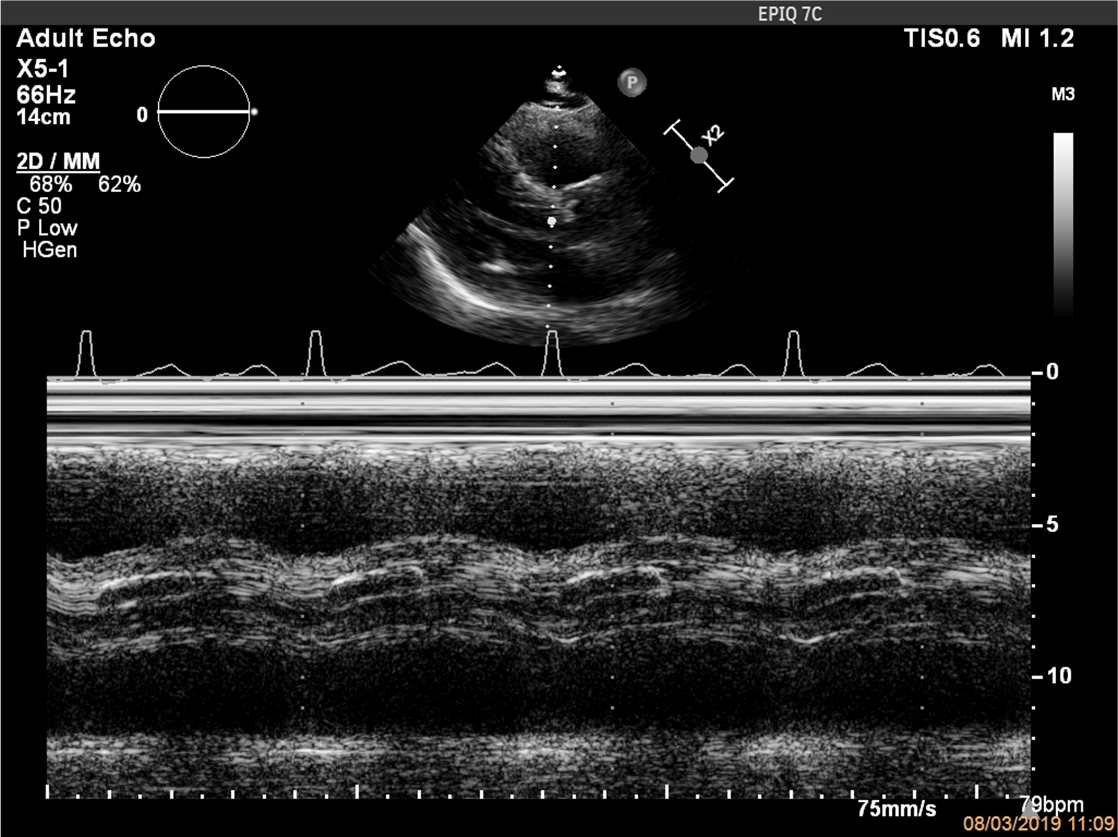
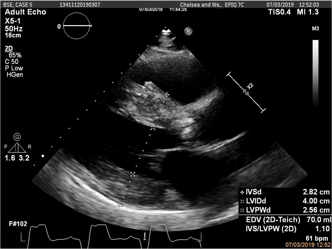
- Based on this M-Mode image, this patient has evidence of:
- Dilated left ventricle
- Restricted aortic valve opening
- Restricted mitral valve opening
- Dilated right ventricle
![]()
![]()
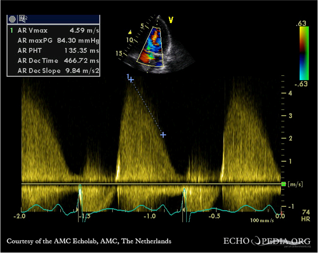
- This patient has evidence of:
- Severe aortic regurgitation
- Restricted aortic valve opening
- Thickened aortic valve leaflets
- Severe MR
![]()
- This patient has evidence of:
- Tricuspid valve
- Bicuspid valve
- Calcified leaflets
- Restricted aortic valve leaflets
![]()
![]()
![]()
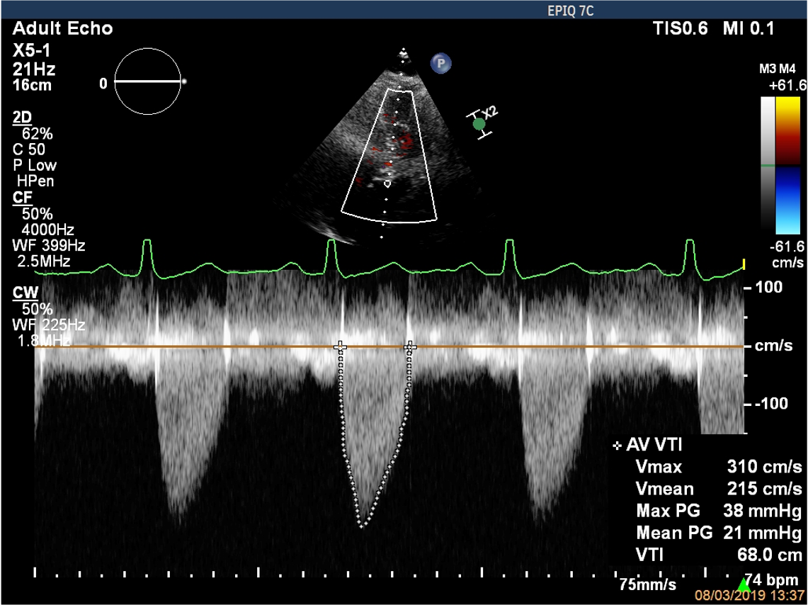
- This patient has:
- Mild aortic stenosis
- Severe aortic stenosis
- Moderate aortic stenosis
- Aortic sclerosis
![]()
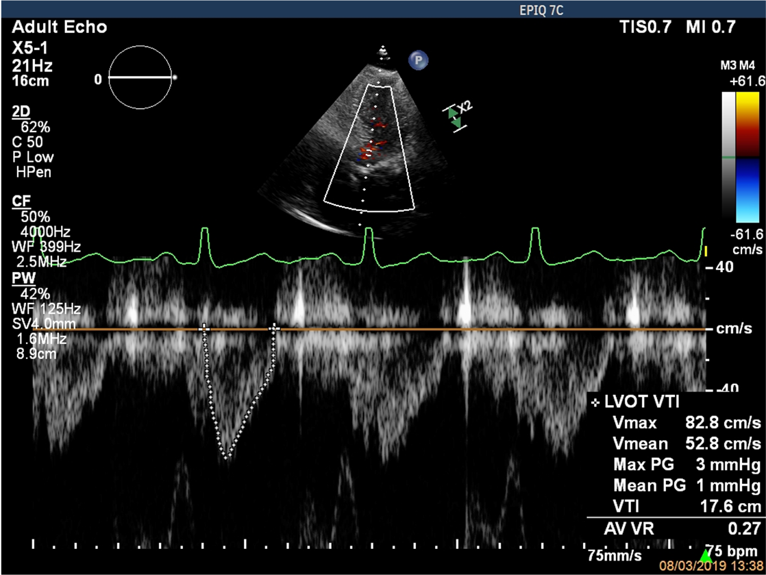
- A DI of 0.27 suggests:
- Mild aortic stenosis
- Moderate aortic stenosis
- Severe aortic stenosis
- Aortic sclerosis
# Case 2
72 year old female with breathlessness
ECG: Normal sinus rhythm
CXR: Unremarkable.
![]()
![]()
- This patient has evidence of:
- Mild AR
- Trivial AR
- Moderate AR
- Severe AS
# Case 3
90 year old male presents with breathlessness
ECG: Low voltage complexes
CXR: Pulmonary oedema.
![]()
![]()
![]()
- This patient has an EF of:
- > 55%
- 40 - 45%
- 35 - 40%
- < 20%
- 30 - 35%
![]()
![]()
- This patient has evidence of:
- Restricted aortic valve leaflets
- Calcified aortic valve leaflets
- Aortic regurgitation
- All of the above
![]()
- There is evidence of:
- Mild AR
- Trivial AR
- Moderate AR
- Severe AR
- No AR
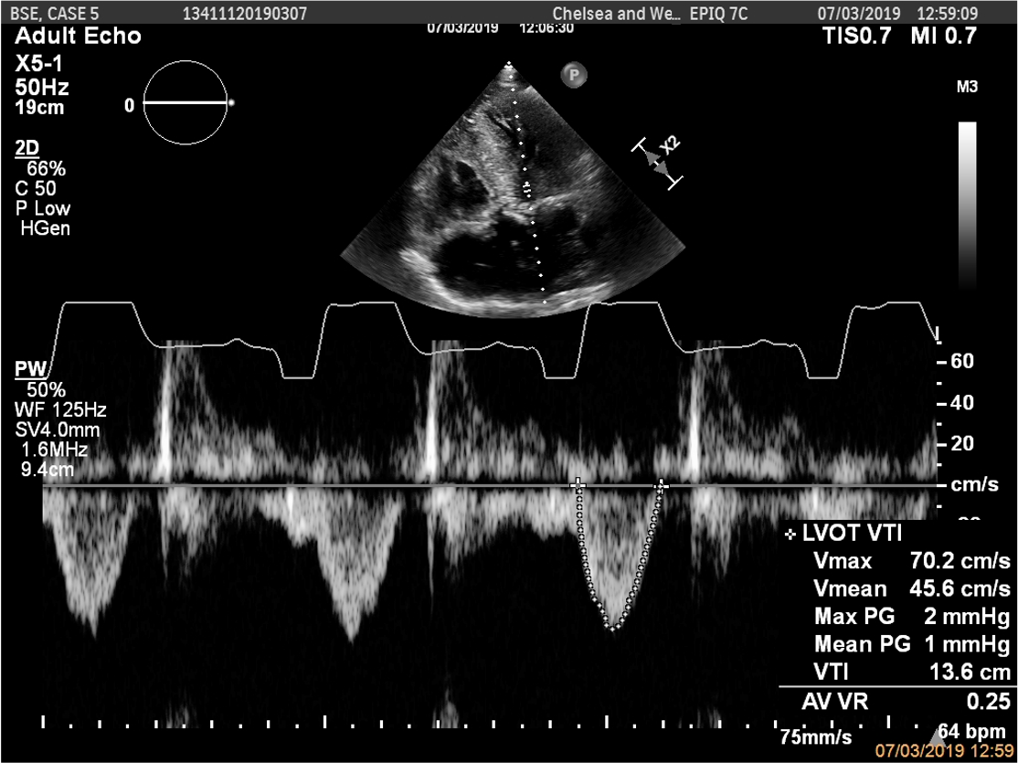
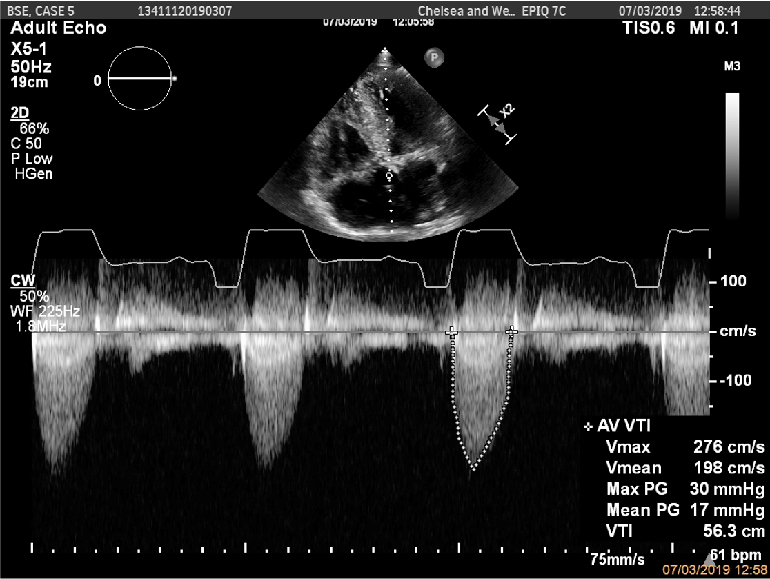
![]()
![]()
- This patient has evidence of:
- Mild AS
- Moderate AS
- Low flow low gradient severe AS
- Paradoxical low flow low gradient severe AS
# Case 4
80 year old female presenting with gross fluid overload, fevers and night sweats
ECG: Normal sinus rhythm
CXR: Pulmonary oedema.
![]()
![]()
- There is evidence of:
- Aortic valve prolapse
- Dilated aortic root
- RCC aortic valve endocarditis
- LCC or NCC aortic valve endocarditis
![]()
- There is evidence of:
- Severe AR
- Severe MR
- Mild MR
- Severe AS
![]()
![]()
- This patient has:
- Mild AR
- Moderate AR
- Severe AR
- No AR
![]()
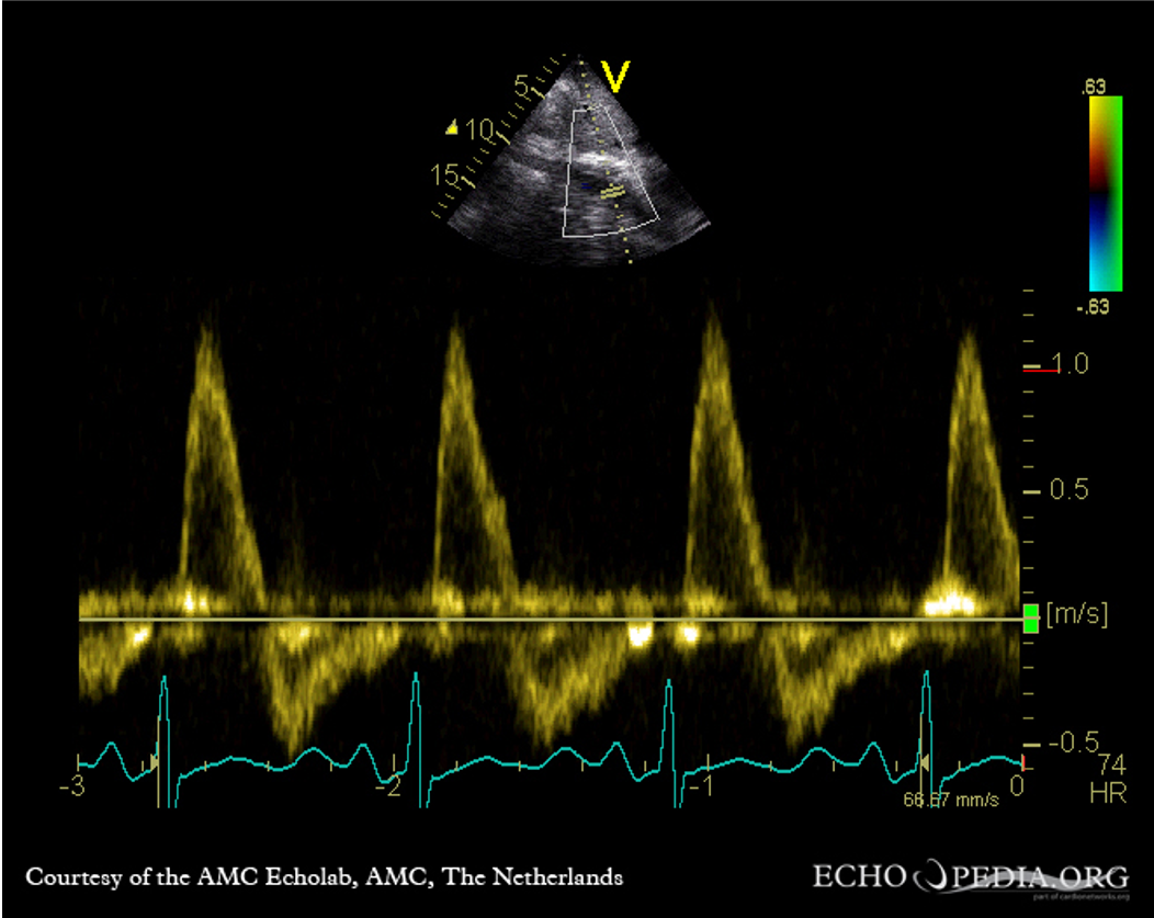
- This pulse wave doppler in the descending aorta confirms the presence of:
- Severe AR
- Normal aortic flow
- Moderate AR
- Co-arctation of the aorta