# Can we always see details well?
我们总是能很好地看到细节吗?
Nickname for echocardiography: Shadow-gram.
超声心动图的昵称:阴影图。
# Limiting factors to taking good images 拍摄良好图像的限制因素
- High body mass index i.e. obese patients
高体重指数,肥胖患者 - Respiratory disease, especially COPD
呼吸系统疾病,尤其是 COPD - Poor patient positioning e.g patient may be in intensive care and not able to be positioned perfectly
患者体位不佳,例如患者可能处于重症监护中,无法完美定位 - Sensitive to pressure on chest
对胸部压力敏感 - Poor technique
技术差 - The pathology may be difficult to see, despite good quality images:
尽管图像质量良好,但以下病理改变可能很难看到:- Left Ventricular (LV) Thrombus
左室血栓 - Inter-atrial Shunts
心房间分流 - High heart rates can make LV wall motion difficult to assess
高心率会使室壁运动难以评估
- Left Ventricular (LV) Thrombus
# The solution 解决方案
- Contrast echocardiography 超声对比剂
- The contrast media utilise gas bubbles
造影剂利用气泡 - Two broad categories: 两大类
- Premanufactured gas mixtures for LV imaging
用于左室成像的预制气体混合物 - Agitated saline or colloid bubbles to see Right-to-Left Shunts
震荡生理盐水或胶体微泡可见右向左分流
- Premanufactured gas mixtures for LV imaging
# Mechanics 机制
- Blood appears black on conventional two dimensional echocardiography, not because blood produces no echo, but because the ultrasound scattered by red blood cells at conventional imaging frequencies is very weak-several thousand times weaker than myocardium 一 and so lies below the displayed dynamic range. [1]
在常规二维超声心动图上,血液呈黑色,不是因为血液不产生回声,而是因为红细胞在常规成像频率下散射的超声非常微弱,比心肌弱几千倍 —— 因此位于所显示的动态范围之下。 - As sound travels from one medium to another, the change in density (known as acoustic impedance) at the interface causes the reflection of sound waves.
当声音从一种介质传播到另一种介质时,交界面密度的变化 (称为声阻抗) 会引起声波的反射。 - The greater the difference in the media densities, the more echogenic the interface.
介质密度的差异越大,交界面回声越强。 - Gas is an excellent contrast agent since it is 100,000 times less dense than blood. [2]
气体是一种极好的造影剂,因为它的密度比血液低 10 万倍。
# LV contrast 左室增强剂
Several brands e.g. Sonovue, Optison, Luminity
几个品牌:声诺维(造影剂,六氟化硫脂质微泡),奥普蒂森(全氟丙烷蛋白质微泡),(全氟丙烷脂质微泡)Given intravenously, followed by a saline flush
静脉注射,然后用生理盐水冲洗
# Sonovue 声诺维
2-8 micrometer size Sulphur Hexafluoride bubbles coated by powder
2-8 微米大小的六氟化硫粉末包裹气泡,干粉制剂Can survive passage through pulmonary circulation.
可以通过肺循环存活下来Mixed with N.Saline and agitated by hand
与生理盐水混合,手动激活0.4 ml injection
0.4 毫升注射液It needs a special setting in the echo machine:
需要在超声机器中进行特殊设置:- LVO 左室显影
# LVO: Left Ventricular Opacification 左室显影
- The power of the ultrasound beam is enough to destroy the bubbles
超声束的能量足以摧毁微泡。 - Continuous imaging can results in loss of contrast effect
连续成像会导致对比剂效果的丧失,通常需要进行 3-4 次对比剂注射 - Mechanical Index (MI) adjustment
机械指数 (MI) 调整- MI = peak negative acoustic pressure/transmitted frequency
MI = 峰值负声压 / 传输频率 - Standard MI = 0.9 - 1.4; lowered to 0.4 or less.
标准 MI 为 0.9-1.4;应用超声增强剂时,选择降至 0.4 或更低来进行成像
- MI = peak negative acoustic pressure/transmitted frequency
# 用途
It delineates the endocardium, making it very easy to see difficult features
它描绘了心内膜,不易观察到的部分变得很容易看到- Typical example: The lateral wall of the LV can be difficult to see in both the Short axis and the 4 Chamber view
典型例子:左室的侧壁在短轴和四腔观中都很难看到 - The use of contrast resolves that issue.
使用对比剂可以解决这个问题
- Typical example: The lateral wall of the LV can be difficult to see in both the Short axis and the 4 Chamber view
LV thrombi (clots) can be difficult to distinguish from artefact.
左室血栓 (血栓) 可能很难与伪像相区别- Ultrasound harmonics can create shadows in the image. These can sometimes look like clot.
超声谐波成像技术,会在图像中产生阴影。这些有时看起来像凝块。 - The use of contrast eliminates this.
使用对比剂可以剔除
- Ultrasound harmonics can create shadows in the image. These can sometimes look like clot.


Excellent for use in LV volume and function by delineating the endocardium, thus making calculations more accurate.
通过描绘心内膜,非常适合用于左室容积和功能的评估,使计算更加准确。![]()
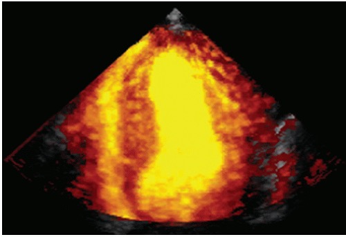
- Power Doppler image recorded at the time of contrast echocardiography using a perfluorocarbon-based agent. Note the excellent signal-to-noise ratio and the marked discrimination between the blood pool and the wall with this imaging method.
使用基于全氟化碳的试剂进行对比超声心动图时记录的功率多普勒图像。注意这种成像方法的出色信噪比和血池与血管壁之间的明显区别。
- Power Doppler image recorded at the time of contrast echocardiography using a perfluorocarbon-based agent. Note the excellent signal-to-noise ratio and the marked discrimination between the blood pool and the wall with this imaging method.
The technique which makes most frequent use of contrast is Dobutamine Stress Echocardiography (DSE).
最常用的造影剂技术是多巴酚丁胺负荷超声心动图 (DSE)。- Ideally, all DSE patients should have LV contrast.
理想情况下,所有 DSE 患者都应应用左室增强剂。
- Ideally, all DSE patients should have LV contrast.
# Emerging indications 新兴适应症
Perfusion studies
心肌灌注研究
3D echocardiography
三维超声心动图

# Limitations 不足
- Expensive 费用
- Occasional allergic reactions
偶尔出现过敏反应
Important point: Echo with LV contrast is much less expensive and time consuming than CMR.
要点:与 CMR 相比,超声 LV 对比剂要便宜得多,耗时也少得多。
# Bubble study 微泡实验(震荡生理盐水成像)
- Used to confirm the presence of a Patent Foramen Ovale (PFO) or Atrial Septal Defect (ASD).
用于确认是否存在卵圆孔未闭 (PFO) 或房间隔缺损 (ASD)- Shunts between the atria can be very difficult to see
心房之间的分流很难看到 - Reason: 原因
- Small 小
- Rely on pressure gradients
分流依赖于心房之间的压力梯度 - Anatomically difficult to obtain good quality images
解剖学上存在困难,如果分流位于心底部,很难获得高质量的图像
- Shunts between the atria can be very difficult to see
# 机制
- The air microbubbles are short-lived and diffuse into the lungs when traversing the pulmonary circulation.
空气微泡是短暂的,当穿过肺循环时会扩散到肺部而被清除,正常情况下不会出现在左心,(如果出现也是少量的,时相靠后的) - Therefore, the microbubbles enter the left heart only in the presence ot a left intracardiac or extracardiac (pulmonary arteriovenous) shunt.
只有在存在左心内或心外 (肺动静脉) 分流的情况下,微泡才会进入左心。 - Saline microbubbles are, therefore, helpful in examining the right heart and identifying shunts. [2:1]
因此,生理盐水微泡有助于检查右心和识别分流。
# Two main categories of mixtures 两大类混合物
8ml N. Saline - 1ml Air - 1ml blood from patient's cannula
8ml 生理盐水 - 1ml 空气 - 1ml 患者的血液(震荡生理盐水)8ml colloid e.g. Gelofusin - 1ml air
8ml 胶体 比如琥珀酰明胶 - 1ml 空气(效果更好)- Limitations 限制
- Colloid gives better image quality but expensive and may give allergic reactions.
胶体可提供更好的图像质量,但价格昂贵,并可能产生过敏反应。
- Colloid gives better image quality but expensive and may give allergic reactions.
- Limitations 限制
The need the use of a 3 way tap and 2 x 10 ml syringes
需要使用三通管和两个 10 ml 注射器Rapid agitation and then injection during a Valsalva manoeuvre to generate R > L atrial pressure gradient upon release.
快速搅拌,然后让患者做 Valsalva 动作来增加右房压力,当右房压力大于左房压力时,如果双房之间有分流,此时静脉注射混合物,微泡会进入左房。

- 一般在心尖四腔切面记录 20 个心动周期。
- 微泡出现的时相,以及出现数量,将帮助判断有没有房间隔缺损,或者卵圆孔未闭。
- 一般心动周期选在第五个或者第六个,如果出现了来自于房间隔部位微泡,一般认为缺损存在。
- 如果是超过了六个心动周期,比如部分微泡有可能来自肺静脉,但这是非常少量的,此时考虑它有可能是经过肺循环没有被清除掉的微泡,通过肺静脉进入到了左侧心脏
- 时相以及部位对判断微泡实验的阴阳性上是非常重要的