# Defining Diastole 定义舒张期
- Diastology 诊断学
- The science and art of characterising left ventricular relaxation, filling dynamics, and their integration into clinical practice.
描述左心室舒张、充盈动力学及其融入临床实践的科学和艺术。
# What is Diastole

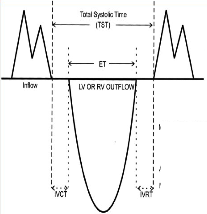
The relationship among LV, LA, and aortic (Ao) pressures and M-mode tracings of the aortic and mitral valve are shown.
显示了 LV、LA 和主动脉(Ao)压力与主动脉瓣和二尖瓣 M 型轨迹之间的关系。Time from aortic valve to mitral valve closure.
从主动脉瓣关闭到二尖瓣关闭的时间,都称为舒张期The isovolumic relaxation time (IVRT) is the interval from aortic valve closure to mitral valve opening. During this interval, LV pressure declines rapidly.
等容舒张时间(IVRT)是从主动脉瓣关闭到二尖瓣打开之间的时间间隔。在此期间,左室压力迅速下降。MV opens when LV pressure < LA pressure
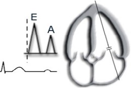
当 LV 压力 < LA 压力时,MV 打开Rapid early filling (E) (~80% OF FILLING)
MV 打开后,左室进行早期的快速充盈 (也就是超声心动图上的 E 波) (约充盈 80%)Diastasis (equalization of pressure b/w atrium and ventricle with little movement of blood)
E 波后续的一段时间为舒张期,此时心室心房压力达到平衡,二尖瓣接近关闭状态,心房和心室间血液流动很少Late diastolic filling 2/2 atrial systole (A) - atrial pressure > vent pressure again, mitral leaflets open again and blood fills LV (20%).
舒张期晚期,充盈 2/2 心房收缩 (就是超声心动图上的 A 波) - 此时心房压力 > 心室压力,二尖瓣叶再次张开,血液充盈左室 (20%)。A rapid rise in LV pressure occurs during the isovolumic contraction time (IVCT), the interval between mitral valve closure and aortic valve opening.
等容收缩时间(IVCT)是从二尖瓣关闭到主动脉瓣打开之间的时间间隔。在此期间,左室压力迅速升高。
RV diastole is similar to LV but duration is shorter.
RV 舒张期与 LV 相似,但持续时间较短。

The relationship between LV volume and the diastolic LV Doppler filling pattern is shown.
显示了 LV 体积与舒张期 LV 多普勒充盈模式之间的关系。Early rapid filling coincides with the E velocity, followed by diastasis (with little or no flow from the LA to the LV), and atrial contraction (which coincides with the late diastolic A velocity).
早期快速充盈与 E 峰速度一致,随后是舒张期 (从左房到 LV 的血流很少或没有血流) 和心房收缩 (与舒张期晚期 A 峰一致)。The Doppler velocity curve, in effect, is the first derivative of the LV volume curve.
多普勒速度曲线实际上是左室容量曲线的一阶导数。Ao, Aortic; MV, mitral valve.
Ideally, the LV is compliant in diastole to fill from a low LA pressure.
理想情况下,LV 在舒张期具有良好的顺应性,较低的 LA 压力即可使左室充盈。LV is stiff in systole to eject the Stroke volume against systolic BP.
左室在收缩期有一定的硬度,这样可以对抗收缩压进行射血。Abnormal LV diastolic function will lead to increased LV diastolic pressure PCWP > 12(10), LVEDP > 16(12).
当左室舒张功能异常,可导致左室舒张压升高,PCWP > 12(10),LVEDP >16(12)。PCWP: pulmonary capillary wedge pressure 肺毛细血管楔压;LVEDP: 左室舒张末压
# Factors influencing LV filling 影响左室充盈的因素
- Active LV relaxation e.g asynchrony; ischaemia
主动左室舒张,如不同步、局部缺血 - Intrinsic passive LV properties
内在被动因素- Myocardial stiffness
心肌僵硬 - Myocardial geometry
心肌几何形状 - Myocardial wall thickness
心肌壁厚度
- Myocardial stiffness
- Extrinsic passive properties
外在被动因素- Pericardial restraint
心包的限制 - Extrinsic compression
外部的压缩 - Ventricular interaction
心室之间的相互作用
- Pericardial restraint
- LA filling pressures
左房充盈压- LA distensibility
左房膨胀性 - LA systole
左房收缩 - Mitral Valve disease
二尖瓣疾病
- LA distensibility
# Diastolic Function Measurements
# Integrated approach
并没有单一的方式能全面评估舒张功能,需要多个方法相结合:
- 2D/M Mode 二维 / M 模式
- LV geometry; 左心室几何形状
- LA size;
- Annular displacement 瓣环漂移
- PW Doppler 脉冲多普勒
- Mitral filling 二尖瓣血流图
- Pulmonary vein inflow 肺静脉血流图
- CW Doppler 连续多普勒
- TR jet to estimate PASP 三尖瓣反流评估肺动脉收缩压
- PR jet to estimate PASP 肺动脉瓣反流评估肺动脉收缩压
- PW tissue Doppler 组织多普勒
- Lateral and medial e' velocities 侧壁和室间隔 e’值
- Colour flow mapping 彩色血流图
- Assess flow velocity propagation 血流传播速度
# 2D/M Mode
Is there LVH?
有没有左室肥厚LV mass index 左室质量指数
What is LA Volume? 左房容积
Abnormal relaxation reduces rate of posterior wall thinning
松弛功能障碍会降低后壁变薄的速率Diminishes rate of LV enlargement in late diastole
也会降低舒张末期左室扩大的速率
| 正常参考值 | |
|---|---|
| LV mass index | > 115g/m2 in men |
| > 95g/m2 in women | |
| LA Volume | > 34ml/m2 |
# LA Volume
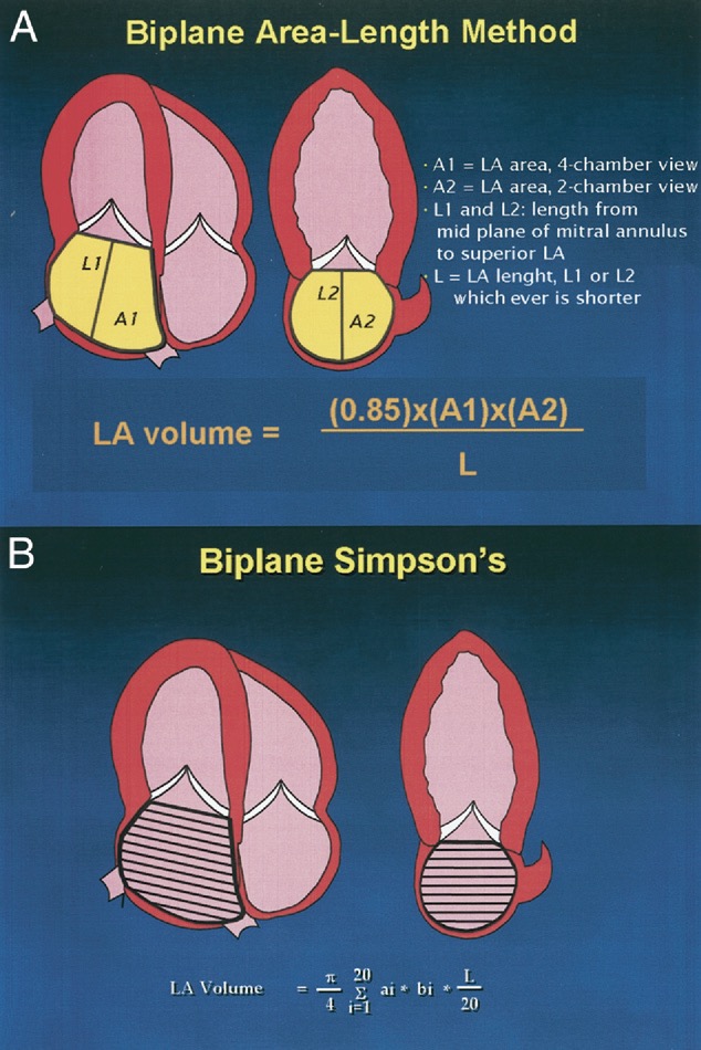
Biplane Methods With Which to Calculate LA Volume.[1]
左房容积反应的是一段时间内,充盈压对左房累积的作用
Barometer of the chronicity of diastolic dlysfunction.
慢性舒张功能障碍的晴雨表LA volume correlates with risk of cardiovascular events including MI, CVA(cerebrovascular accident), A fib and CHF.
LA 容量与心血管事件风险相关,包括心梗、脑卒中、房颤和慢性心衰等More stable than LV filling pressures.
比 LV 充盈压更稳定。
# PW Doppler
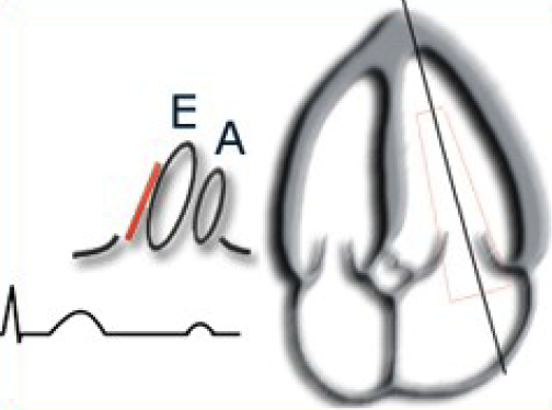
# Transmitral Doppler Inflow 经二尖瓣血流频谱

- Sample at leaflet tips 在瓣叶尖取样
- Must be precise 必须精确
- Sweep speed 50-100mm/s
扫描速度 - Correlate with ECG
与心电图相关,E 波在 t 波之后,A 波在 p 波之后


- Normal pattern of left ventricular diastolic filling.[2]
左室舒张充盈正常模式。 - Pulsed Doppler recording in an apical four-chamber view with the sample volume positioned at the mitral leaflet tips (left) is used to measure E velocity, A velocity, and the deceleration time (arrow).
在心尖四腔视图中脉冲多普勒记录,并放置取样容积放在二尖瓣叶尖 (左) 用于测量 E 峰速度,A 峰速度和减速时间 (箭头)。 - The flow signal recorded with the sample volume positioned at the level of the mitral annulus level (right) is used for measurement of atrial flow duration.
用位于二尖瓣环水平 (右) 的取样容积记录的血流信号用于测量心房血流持续时间。 - If transmitral stroke volume is calculated, the annular flow signal is used for the velocity-time integral.
如果计算二尖瓣搏出量,则使用环状血流信号进行速度 - 时间积分。 - DFP, Diastolic filling period; DT, deceleration time; IVRT, isovolumic relaxation time; VTI, velocity-time integral.
DFP,舒张充盈期;DT,减速时间;IVRT,等容松弛时间;VTI,速度 - 时间积分。

- Do not confuse EDT(E wave deceleration time) and PHT(pressure half time)
不要混淆 E 波减速时间和压力减半时间 - DT 减速时间,E 波的峰值降到最低点的时间

- Raised mid diastole velocities may Suggest delayed relaxation.
舒张中期速度升高可能表明舒张延迟。

- The isovolumic relaxation time (IVRT) is measured from aortic valve closure and the onset of mitral flow (arrows) and measures 96 ms in this example.
等容舒张时间。主动脉瓣关闭后二尖瓣打开之前,Normal: 70-90 ms. - 取样束放置在偏向左室流出道的位置,既可以描述主动脉瓣射血的时间,也可以描述二尖瓣瓣口血流的时间
| Normal PW Measurement | Age group (y) | |||
|---|---|---|---|---|
| 16-20 | 21-40 | 41-60 | > 60 | |
IVRT (ms) | 50 ± 9 | 67 ± 8 | 74 ± 7 | 87 ± 7 |
E/A ratio | 1.88 ± 0.45 | 1.53 ± 0.40 | 1.28 ± 0.25 | 0.96 ± 0.18 |
EDT (ms) | 142 ± 19 | 166 ± 14 | 181 ± 19 | 200 ± 29 |
A duration (ms) | 113 ± 17 | 127 ± 13 | 133 ± 13 | 138 ± 19 |
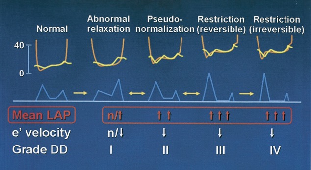
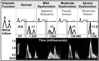
Normal | Abnormal relaxation | Pseudo-normal filling pattern(PNF) | Restrictive filling pattern | ||
|---|---|---|---|---|---|
| reversible 可逆 | irreversible 不可逆 | ||||
| E/A ratio | 1-2 | < 0.8 | E/A 或 DT 很难与正常相区分,需要结合其他指标来区分舒张期功能。比如组织多普勒的 e' 出现降低,E/e' 升高到>15 的不正常值。 | > 2,在 E/A 比率上有所区分,不可逆的 E/A 远大于 2 | |
| DT(ms) | 150-200 | > 200, 延长 | < 160, 缩短 | ||
| IVRT(ms) | 50-100 | > 100, 延迟 | < 80, 缩短 | ||
所有舒张功能受损的患者,都会导致左房压力增高
# Pulmonary Venous Doppler Flow 肺静脉血流频谱图
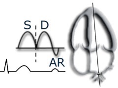
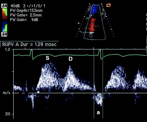
- 可以反应左室的舒张功能,心尖四腔,取样容积放在肺静脉 0.5-1cm 进行频谱图描绘[3]
| 4 phase | |||
|---|---|---|---|
| S wave | 收缩期 | LA relaxation | 左房舒张 |
| LV annulus displacement | 左室瓣环移位 | ||
| D wave | 舒张期前向血流 | Ventricular relaxation | 心室舒张 |
| Ar wave | 舒张期晚期逆向血流 | Atrial contraction | 心房收缩 |
- Main measurements
- S velocity
S 波速度 - D velocity
D 波速度 - S/D ratio
S/D 比率 - Ar Velocity
Ar 波返流速度
有时写为 a 波,以和二尖瓣血流频谱的 A 波区分
- S velocity
| Measurement | ||
|---|---|---|
| S/D ratio | Normally | > 1 |
With raised LVEDP becomes | < 1 | |
| Ar Velocity | As LVEDP size and duration will increase (cf A wave duration with DECREASE) | |
当左室舒张末压升高时,Ar 的流速和持续时间都会增加 (而 A 波持续时间则减少) | ||
Careful with influencing factors on PV flow 注意以下影响因素
- Advanced age 高龄
- Age will increase S/D ratio; Ar max and Ar duration
年龄会增加 S/D 比率;Ar max 和 Ar 持续时间
- Age will increase S/D ratio; Ar max and Ar duration
- Tachycardia (SD fusion) 心动过速(SD 融合)
- First degree AV block I 度房室传导阻滞
- MR 二尖瓣反流
- Raise Preload with increase S 增加的前负荷
- LV systolic dysfunction 左室收缩功能障碍
- Advanced age 高龄
Pulm vein flow velocity Tips/Tricks 测量技巧
- Use sweep speeds of 100mm/s 扫描速度
- Perform with apnoea 呼吸暂停
- MR may annul or even reverse S velocity
二尖瓣反流可以抵消甚至反转 S 速度- Hence do not use if there is significant MR
因此,如果有明显的 MR,请不要测量
- Hence do not use if there is significant MR
- Do not use immediately after AF DCCV(direct current cardioversion) due to atrial stunning
由于心房顿抑,不要在房颤直流电复律后立即测量 - Be wary of relative degrees of pulm vein stenosis if the patient has had a RF(radiofrequency) AF ablation in past.
如果患者过去做过房颤射频消融术,要注意肺静脉狭窄的相对程度。
# Tissue Doppler Imaging (e') 组织多普勒
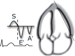
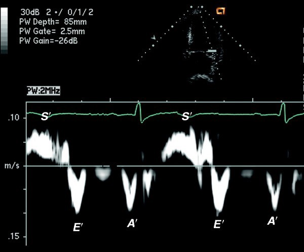
心尖四腔,取样线放置在二尖瓣瓣环处,进行瓣环运动速度的描绘
可以得到
- 收缩期 s' 波
- 舒张早期 E' 波,与经二尖瓣血流频谱图中的 E 波出现时相一样,在 t 波之后
- 舒张晚期 A' 波,出现在 p 波之后
E/e' ratio (combo of transmitral flow velocity and annular velocity) is best parameter for predicting mean LVEDP (< 8 nl, > 15 abnl); but cannot be used in isolation!
E/e' 比率 (跨二尖瓣血流速度和二尖瓣环运动速度的组合) 是预测平均左室舒张末压 (<8nL,>15abnl) 的最佳参数,但不能单独使用!This is the best correlate with NORMAL LVEF and no mitral annular issue.
射血分数正常以及无二尖瓣瓣环问题时,E/e' 比率有很好的相关性
| Measurement | ||
|---|---|---|
| E/e' ratio | normal | < 8 |
| abnormal | > 15 | |
- Tips with TDI 注意事项
- Mitral annular calcification or a mitral annular ring will reduce velocities
二尖瓣环钙化或二尖瓣环植入,会减慢运动速度,减低 e' 值 - Reduced LVEF, especially local infarction will lead to inaccurate assessment
左室射血分数降低,尤其是取样线处有局部梗死,将导致评估不准确 - e' will be increased with significant degrees of primary MR
e' 在显著的原发性二尖瓣反流时出现增高 - Reduced e' is consistent across all degrees of diastolic dysfunction and is an early marker; however also a normal part of ageing.
所有程度的舒张功能障碍中 e' 都是降低的,因此 e' 的下降可作为舒张功能障碍的早期标志;但随着年龄的增长,e' 也会出现下降。
- Mitral annular calcification or a mitral annular ring will reduce velocities
# Mitral inflow Propagation Velocity (Vp) 二尖瓣血流传播速度

Propagation velocity (dashed line) recorded with the color Doppler M-mode cursor positioned in the center of the mitral annulus in an apical four-chamber view.
在心尖四腔切面下,用位于二尖瓣环中央的彩色多普勒 M 型光标记录传播速度 (虚线)。The slope of the Doppler flow as it moves from the annulus (bottom of scale) to the apex (top of the scale) reflects the rate of LV relaxation.
当多普勒血流从环部 (标尺底部) 移动到心尖 (标尺顶部) 时,其斜率反映了左室舒张的速度。Colour M mode 100mm/s 彩色 M 模式
Shift colour flow baseline down so highest velocity is blue.
将彩色血流基线向下移动(低于奈奎斯特极限),使最高流速为蓝色,并描绘,得到斜率来反映血流传播速度Slope relates to relaxation
斜率与松弛有关临床非常规,了解
| Measurement | ||
|---|---|---|
| Vp | > 55cm/s | normal |
| E/Vp | > 2.5 | correlates with raised LVEDP 左室舒张末压升高 |
# Global strain rate 整体应变率
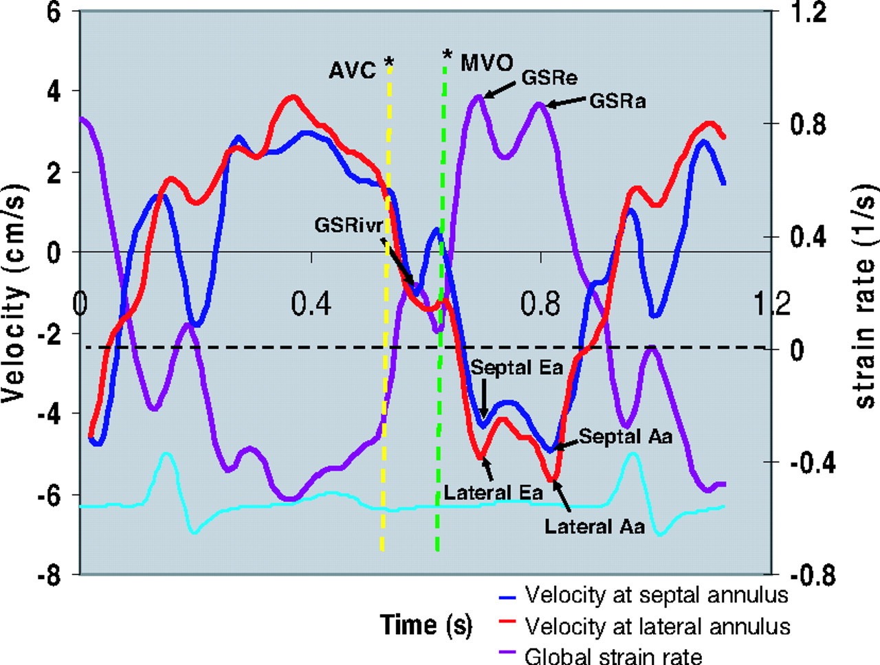
- 也可以用左室舒张功能的评估
- 了解[4]
# Diastolic Function Grades
| Decel Time | 140-250ms | |
| E/e' | < 8 | normal |
| E/e' | > 15 | LVEDP > 18mmHg |
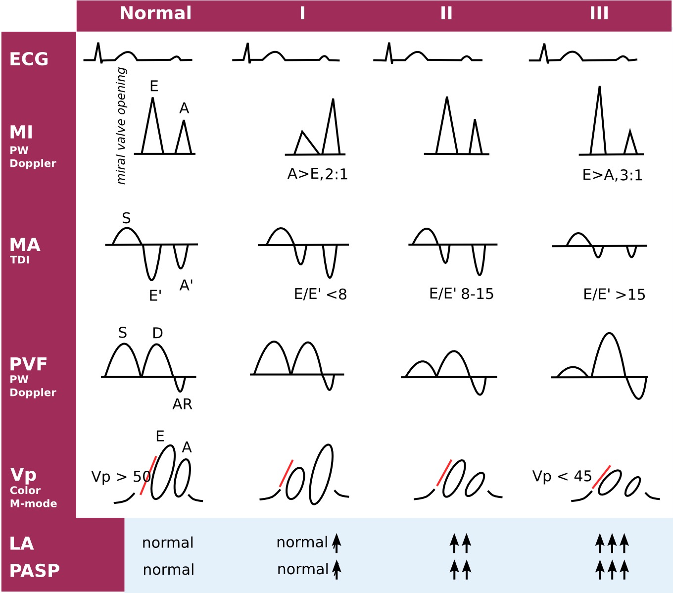
- I: impaired relaxation 松弛受损
- II: moderate diastolic dysfunction (pseudonormal) 中度舒张功能障碍(假性正常)
- III: restrictive left ventricular filling (impaired LV compliance) 限制性左心室充盈(LV 顺应性受损)
- ECG: electrocardiogram, MI: mitral inflow, MA: mitral annular velocities, PVF: pulmonary venous flow, Vp: velocity of flow progression, LA: left atrium, PASP: pulmonary artery systolic pressure
ECG:心电图,MI:二尖瓣流入,MA:二尖瓣环速度,PVF:肺静脉流量,Vp:速度流动进展,LA:左心房,PASP:肺动脉收缩压。
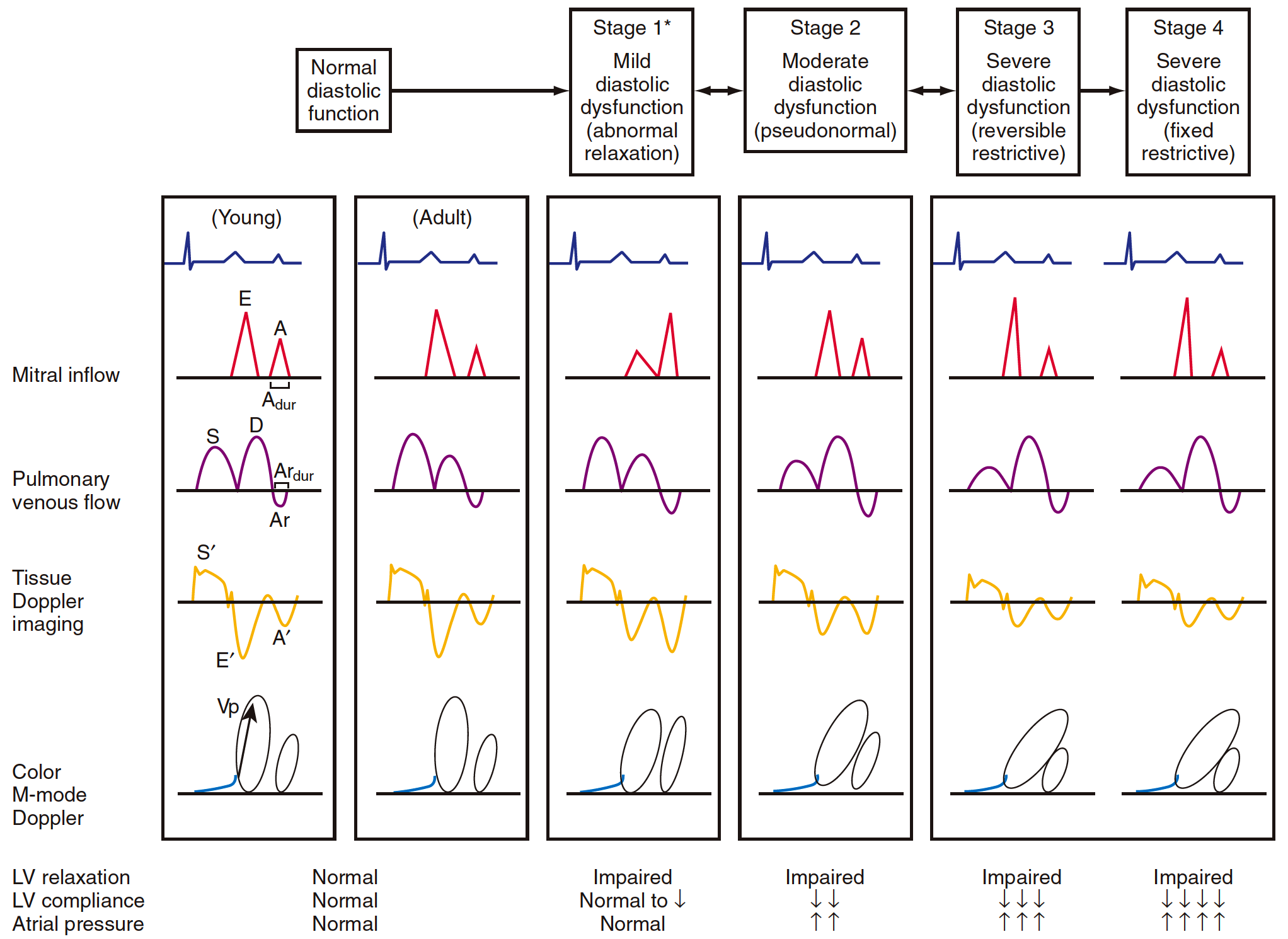
- Stages of diastolic function. 舒张功能的阶段。
- Schematic representation of the typical patterns seen with mitral inflow, pulmonary venous flow, tissue Doppler echocardiography, and color M-mode propagation velocity (Vp) for normal (young and adult), impaired relaxation, pseudonormal, and restrictive diastolic function.
二尖瓣血流、肺静脉血流、组织多普勒超声心动图和彩色 M 型传播速度 (Vp) 在正常 (年轻人和成人)、松弛受损、假性正常和舒张期功能受限时的典型模式示意图。 - The stages of diastolic dysfunction can be determined using an integrated approach with these four different modalities for assessment of diastolic flow pattern.
评价舒张期血流频谱的四种不同方法综合起来,可以确定舒张期功能障碍的分期。 - A, Late mitral filling;
- A′, tissue Doppler atrial velocity;
- Ar, pulmonary vein atrial reversal velocity;
- Ardur, duration of pulmonary vein Ar;
- D, pulmonary vein diastolic filling;
- E, early mitral filling;
- E′, tissue Doppler early diastolic velocity;
- S, pulmonary vein systolic filling;
- S′, tissue Doppler systolic velocity;
- Vp, propagation velocity.
- Schematic representation of the typical patterns seen with mitral inflow, pulmonary venous flow, tissue Doppler echocardiography, and color M-mode propagation velocity (Vp) for normal (young and adult), impaired relaxation, pseudonormal, and restrictive diastolic function.
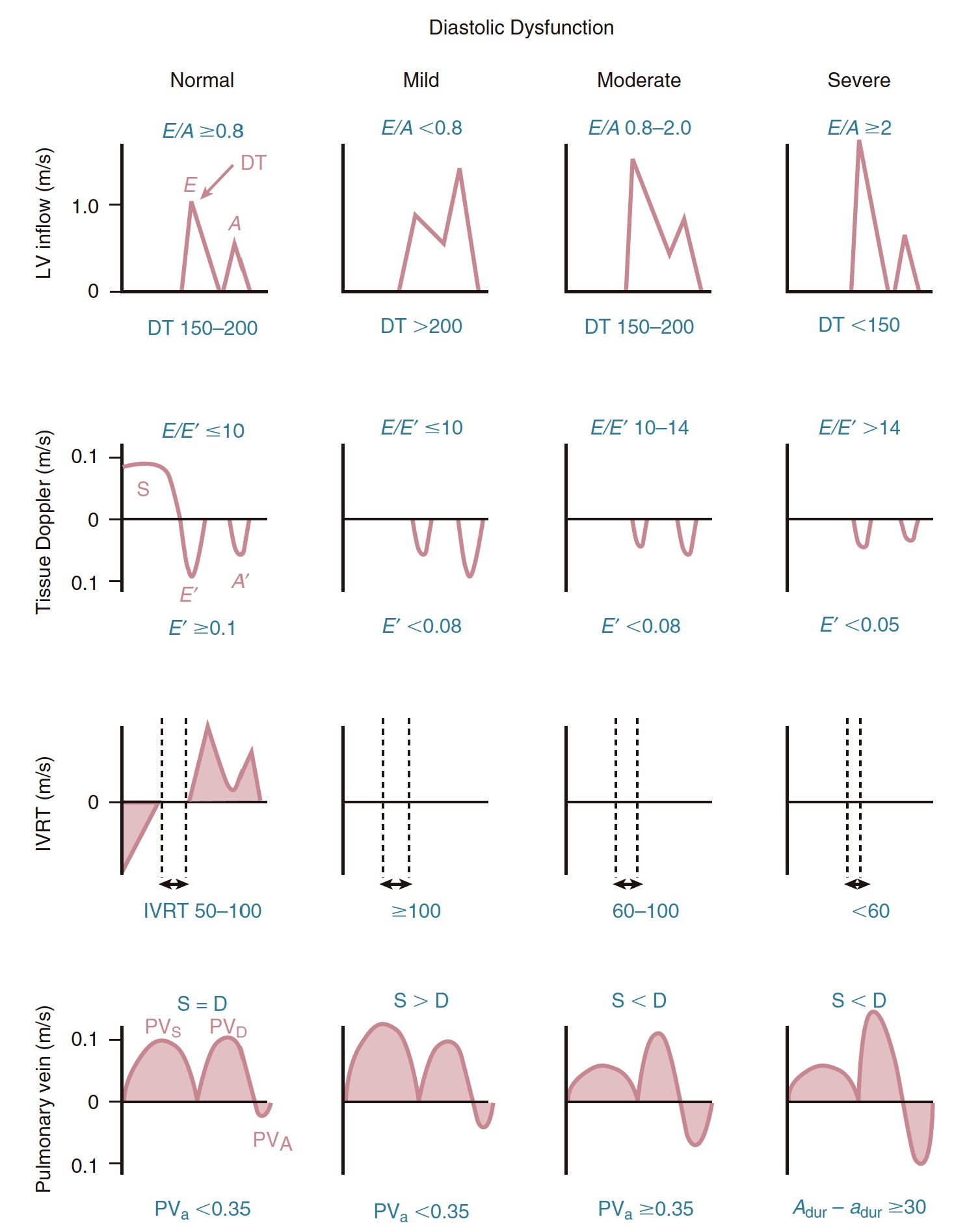
- Doppler findings in patients with normal diastolic function and with mild, moderate, and severe diastolic dysfunction. 舒张功能正常以及轻度、中度和重度舒张功能障碍患者的多普勒表现。
- The top row shows LV inflow with early (E) and atrial (A) phases of diastolic filling, the second row from the top shows tissue Doppler imaging recorded at the septal side of the mitral annulus with the myocardial early (E′) and atrial (A′) velocities and the expected ratio of (E/E′), the third row from the top shows the isovolumic relaxation time (IVRT), and the bottom row shows the pulmonary venous inflow pattern with systolic (S) and diastolic (D) antegrade flow and the pulmonary vein atrial (PVa) reversal of flow.
第一排显示二尖瓣环左室流出道的舒张期充盈早期 (E) 和心房 (A) 相,倒数第二排显示二尖瓣环间隔侧心肌早期 (E‘) 和心房 (A’) 的血流速度和预期的比值 (E/E‘),倒数第三排显示等容舒张期 (IVRT),最下面一排显示肺静脉流入模式,收缩期 (S) 和舒张期 (D) 顺行血流,肺静脉 (PVA) 血流逆转。
# Approch to the Patient 患者评估

When LV size, wall thickness, and ejection fraction (EF) are normal, further evaluation of diastolic function is needed only in the presence of LA enlargement or an abnormal E/A ratio for age.
当 LV 大小,壁厚和射血分数 (EF) 正常时,仅在 LA 增大或年龄 E/A 比率异常的情况下,才需要进一步评估舒张功能。In patients with ventricular hypertrophy or dilation with a normal ejection fraction, diastolic function should be fully evaluated, particularly if clinical concern exists that diastolic dysfunction is the cause of symptoms.
对于射血分数正常的心室肥大或扩张患者,应全面评估舒张功能,尤其是当临床关注舒张功能障碍是症状的原因时。When ejection fraction is reduced, the first step is to evaluate for elevated filling pressures.
当射血分数降低时,第一步是评估是否有较高的充盈压力。If simple criteria for elevated filling pressures are not present, a more complete evaluation of diastolic function is appropriate.
如果不符合充盈压力升高的简单标准,则应对舒张期功能进行更全面的评估。DT, Deceleration time; IVRT, isovolumic relaxation time; LVH, LV hypertrophy; PVD, peak diastolic velocity; PVS, peak systolic velocity.
根据左室的大小、室壁厚度和 EF 值进行不同的评估
二尖瓣的血流频谱图和组织多普勒是必须描绘的,尽量区分舒张功能障碍的严重程度
如果血流频谱和组织多普勒无法评估,可以加用 IVRT 和 肺静脉血流频谱
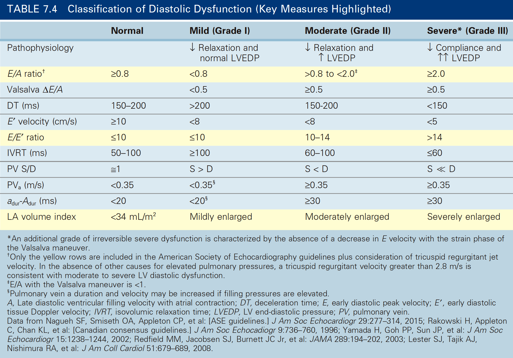
# Presence and severity of diastolic dysfunction
| Normal | Mild (Grade I) | Moderate (Grade II) | Severe* (Grade III) | |
|---|---|---|---|---|
| Pathophysiology | ↓Relaxation and normal LVEDP | ↓Relaxation and ↑LVEDP | ↓Compliance and ↑↑LVEDP | |
| E/A ratio | ≥ 0.8 | < 0.8 | > 0.8 to <2.0 | ≥ 2.0 |
| Valsalva △E/A | < 0.5 | ≥ 0.5 | ≥ 0.5 | |
| DT (ms) | 150-200 | > 200 | 150-200 | < 150 |
| E’ velocity (cm/s) | ≤ 10 | < 8 | < 8 | < 5 |
| E/E’ ratio | ≤ 10 | ≤ 10 | 10-14 | > 14 |
| IVRT (ms) | 50-100 | ≥ 100 | 60-100 | ≤ 60 |
| PV S/D | ≈1 | S > D | S < D | S ≪ D |
| PVₐ(m/s) | < 0.35 | < 0.35 | ≥ 0.35 | ≥ 0.35 |
| a_dur_ - A_dur_ (ms) | < 20 | < 20 | ≥ 30 | ≥ 30 |
| LA volume index | < 34 mL/m2 | Mildly enlarged | Moderately enlarged | Severely enlarged |
# Assessment of LVEDP
在左室舒张末压升高的情况下,各指标的参考值
| LA | E/A | EDT | Ar | Ar dur - A dur | S/D | DDT | E/e' | E/Vp |
|---|---|---|---|---|---|---|---|---|
| ↑ | >2 | < 150ms | > 0.35 m/s | >30 ms | S < D | <175 ms | > 15 | >2 |
Lester, S. J. et al. Unlocking the mysteries of diastolic function: deciphering the Rosetta Stone 10 years later. J Am Coll Cardiol 51, 679–689 (2008). ↩︎
Catherine M. Otto MD. Textbook of Clinical Echocardiography ↩︎
https://echobasics.de/diastole-en.html ↩︎
Wang, J., Khoury, D. S., Thohan, V., Torre-Amione, G. & Nagueh, S. F. Global diastolic strain rate for the assessment of left ventricular relaxation and filling pressures. Circulation 115, 1376–1383 (2007). ↩︎