# Basic views of the right ventricle
# RV dimensions and structure 右室大小与结构
# RV Inflow Dimensions
- Assessed in apical 4 chamber
- Base (RVD1): below the TV annulus < 4.2cm
右心室基底部内径 - Mid (RVD2): mid-RV < 3.5cm
中部 - Length (RVD3): base to apex < 8.6cm
右心室长径
- Base (RVD1): below the TV annulus < 4.2cm
# RV Outflow Dimensions
- Parasternal long axis 胸骨旁长轴
- RVOT Prox in PLAX: < 3.1cm
右室流出道近端内径
- RVOT Prox in PLAX: < 3.1cm
- Parasternal short axis 胸骨旁短轴
- RVOT Prox at AV level: < 3.6cm
右室流出道近端内径 - RVOT distal at PV level: < 2.8cm
右室流出道远端内径 - PA diameter: < 2.3cm.
肺动脉内径
- RVOT Prox at AV level: < 3.6cm
# RV Dilatation 右室扩张
Causes of RV Dilatation
- Pressure overload 压力负荷过高
- Volume overload 容量负荷过高
- Myocardial Disease 心肌病
Qualitative 定性的
- Assessed in the apical four chamber view
在心尖四腔切面中评估 - The RV should be less than two thirds tje size of the LV
正常情况下,4C 下 RV 的尺寸应小于 LV 的三分之二 - Should not from part of the apex.
正常情况下,4C 下看不到右室的心尖部分
- Assessed in the apical four chamber view

# Eccentricity Index (EI) 偏心指数

D1 = Minor axis dimension perpendicular to septum
垂直于室间隔的短轴尺寸D2 = Minor axis dimension perpendicular to D1.
垂直于 D1 的短轴尺寸A normal round shape would yield a ratio of 1.0, whereas septal flattening would result in an eccentricity index greater than 1.0.
正常的圆形将产生 1.0 的比率,而室间隔变平将导致偏心指数大于 1.0。EI cutoff > 1.0
- If EI > 1 in systole it suggests a pressure overload state
发生在收缩期时,表示压力超负荷状态 - If EI > 1 in diastole it suggests a volume overload state
发生在舒张期时,表示容量超负荷状态
- If EI > 1 in systole it suggests a pressure overload state
| EI 值 | ||
|---|---|---|
| ≤ 1 | Normal | |
| > 1 | Systole and Diastole | RV Pressure Overload |
| Diastole | RV Volume Overload | |


# Pressure Overload 压力负荷过高
Septum flattens in systole and diastole.
观察室间隔的状态,室间隔在收缩和舒张期变得扁平。The eccentricity index(EI) can quantify this.
偏心指数可以量化这一点。Causes 病因
- Pulmonary hypertension 肺动脉高压
- Acute pulmonary embolism 急性肺栓塞
- RV outflow tract obstruction 右室流出道梗阻


# Pulmonary Hypertension 肺动脉高压
Five groups: 分为五个类型
- Pulmonary artery hypertension (PAH) 原发性肺动脉高压
- Due to left heart disease 左心疾病所致
- Due to lung disease/hypoxia 肺部疾病 / 缺氧所致
- Chronic thromboembolic PH (CTEPH) 慢性血栓栓塞性肺动脉高压
- Due to unclear aetiology 病因不明
需要评估肺动脉收缩压和舒张压
Evaluated during routine transthoracic echocardiography using tricuspid or pulmonary doppler and estimating right atrial pressure using the IVC.
在常规经胸超声心动图中,肺动脉收缩压可以根据三尖瓣反流的跨瓣压差以及右房压来评估,或者用肺动脉多普勒成像来评估肺动脉压力情况,并使用下腔静脉评估右房压力。Calculating PA Systolic Pressure
(in the absence of RVOT obstruction)
(在没有右室流出道阻塞的情况下)


# Acute Pulmonary Embolism 急性肺栓塞
- Can cause RV dilatation and impaired systolic function
会导致 RV 扩张和收缩功能受损 - RV pressure overload (EI > 1 in systole)
RV 压力负荷过大 (收缩期 EI> 1) - McConnell's sign is seen in central clots - Akinesia of the mid free wall but normal motion at the apex.
麦康奈尔征见于中央型血栓 -- 右室呈明显扩张状态,心尖部保持正常运动和收缩,右室游离壁中段呈现运动迟缓或无运动状态
# Volume Overload 容量负荷过高
Causes
- Atrial septal defect, partial anomalous pulmonary venous drainage, coronary sinus defect
从左到右的心内分流:房间隔缺损、部分型肺静脉异位回流、冠状静脉窦型缺损 - Tricuspid valve disease
三尖瓣疾病,明显的重度三尖瓣反流 - Ebstein anomaly
埃勃斯坦畸形 - Pulmonary regurgitation
肺动脉瓣反流 - RV outflow tract obstruction.
右室流出道梗阻
- Atrial septal defect, partial anomalous pulmonary venous drainage, coronary sinus defect
The septum flattens during diastole. The eccentricity index can quantify this.
胸骨旁短轴,乳头肌水平,静止帧观察收缩期、舒张期。室间隔在舒张期变平。偏心率指数可以量化这一点。
# Atrial septal defect 房间隔缺损
- Initially left to right shunt - Leads to increased pulmonary flow and pulmonary hypertension
最初左向右分流 - 导致肺循环血流量增加和肺动脉高压 - Leads to dilated right atrium and right ventricle
导致右心房和右心室扩张 - Eisenmenger's syndrome: Shunt reversal (right to left) due to PA pressure > LA pressure.
病情进一步进展可引起艾森曼格综合征:由于 PA 压力>LA 压力导致分流逆转(从右向左)。

- Upper sinus venosus defect 上腔型缺损
- Lower sinus venosus defect 下腔型缺损
- Secundum defect 中央型缺损(最常见)
- Involving coronary sinus 静脉窦型缺损
- Primum defect 原发孔缺损
- Coronary sinus defect 冠状窦缺损
Results in blood flow from the left atrium to the right atrium and hence RA, RV and RVOT dilatation.
导致血液从左心房流向右心房,从而导致 RA、RV 和 RVOT 扩张。
# Bubble Echocardiography 微泡超声心动图 / 震荡生理盐水成像
- Agitated saline injected peripherally
外周注射震荡生理盐水 - Bubble diameter 50-90um
气泡直径 50-90 微米 - Destroyed as they pass through pulmonary capillaries (unless there is a pulmonary shunt)
当它们通过肺毛细血管时被破坏 (除非有肺分流) - Can diagnose PFO, ASD, persistent left superior vena cava.
可诊断卵圆孔未闭、房间隔缺损、永存左位上腔静脉。
# Anomalous Pulmonary Venous Drainage 肺静脉异位回流
Partial: One or two of the pulmonary veins drain into the RA Or a vessel instead of the LA
部分型:一条或两条肺静脉流入 RA 或血管,而不是 LA- Can be associated with an ASD
可与 ASD 相关 - Diagnosed in older age with RA/ RV dilatation and PH
老年患者被诊断为 RA/RV 扩张和 PH
- Can be associated with an ASD
Total: All of the pulmonary veins drain blood into a vessel or chamber other than the left atrium
完全型:所有肺静脉都将血液排入左心房以外的血管或腔内- Patent PFO, PDA or ASD must be present or this condition is fatal.
必须存在 PFO、PDA 或 ASD,否则这种情况是致命的。
- Patent PFO, PDA or ASD must be present or this condition is fatal.
由于超声心动图的局限性,很难观察到,仅仅能看到右心扩张的情况;此时需要借助磁共振或者 CT 进行诊断。
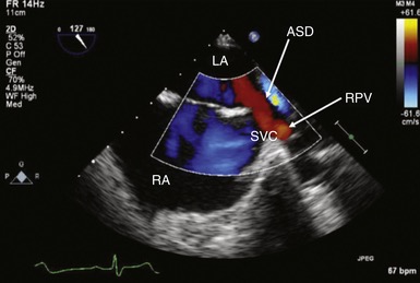
Right pulmonary vein draining directly into the SVC - note the associated ASD.
右肺静脉直接引流至上腔静脉 - 注意相关的房间隔缺损。Superior sinus venosus defect. Midesophageal bicaval view: this type of atrial septal defect (ASD) is mostly associated with a partial anomalous pulmonary vein return. In red, inflow of right ventricle (RV) into superior vena cava (SVC). LA, Left atrium; RA, right atrium; RPV, right pulmonary vein.
上窦静脉缺损。食管中段双腔观察:这种类型的房间隔缺损 (ASD) 主要与部分异常的肺静脉回流有关。红色,右心室 (RV) 流入上腔静脉 (SVC)。LA,左心房;RA,右心房;RPV,右肺静脉。
# Tricuspid Regurgitation 三尖瓣反流
- Causes: functional due to RV dilatation, carcinoid, infective endocarditis, pacing leads
右室扩张、类癌、感染性心内膜炎、起搏电极所致的功能性反流 - Chronic TR results in RV volume overload and hence dilatation
慢性 TR 导致右室容量超负荷,从而导致右室扩张 - Can estimate the pulmonary pressure using TR max gradient + right atrial pressure.
可以使用连续多普勒测 TR 最大梯度 + 右心房压力来估计肺动脉压。
# Ebstein Anomaly
- Congenital malformation
先天性畸形 - Downward displacement of tricuspid valve (apical displacement of the septal leaflet by at least 8mm)
三尖瓣向下移位 (隔叶朝心尖移位至少 8 mm) - Dilated RA due to significant TR
严重的 TR 导致 RA 扩张 - Atrialisation of the upper RV due to displaced tricuspid valve
三尖瓣移位导致 RV 上部心房化 - Dilated RV with reduced function.
右心室扩张,功能减退。
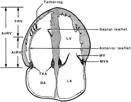
- Schematic of anatomic abnormalities in Ebstein anomaly. AnRV, anatomic right ventricle; AtRV, atrialized right ventricle; FRV, functional right ventricle; MVA, mitral valve annulus; TVA, tricuspid valve annulus.
Ebstein 畸形的解剖异常示意图。ANRV,解剖右室;AtRV,房化右室;FRV,功能右室;MVA,二尖瓣环;TVA,三尖瓣环。
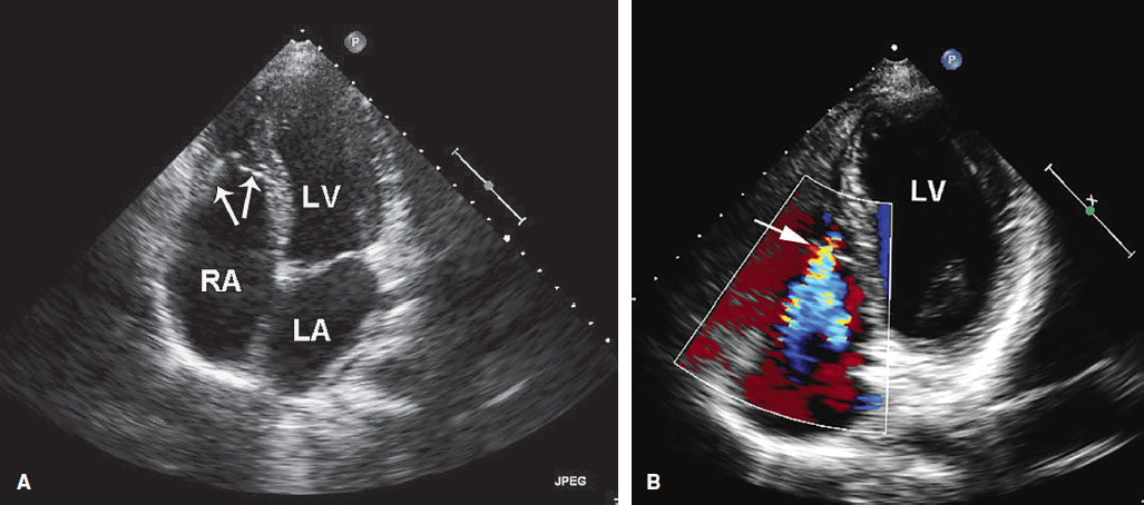
- An extreme example of Ebstein anomaly. From the apical four-chamber view (A), the tricuspid valve leaflets (arrows) are displaced far into the right ventricular apex. In B, note the origin of the tricuspid regurgitation jet far into the right ventricular cavity (arrow).
Ebstein 畸形的一个极端例子。从心尖四腔切面 (A),三尖瓣瓣叶 (箭头) 移位到右室心尖部。在图 B 中,注意三尖瓣返流的起始处远进入右室腔 (箭头)。
# Pulmonary Regurgitation 肺动脉瓣反流
- Assessed in parasternal short axis or parasternal RV outflow
在胸骨旁短轴或胸骨旁右室流出道评估,在肺动脉水平放置彩色多普勒 - Usually due to ring dilatation due to chronic pulmonary hypertension
通常是由于慢性肺动脉高压引起的环状扩张 - Or pulmonary artery dilatation due to connective tissue disorders
或由于结缔组织疾病引起的肺动脉扩张 - RV dilatation and impaired systolic function
右室扩张与收缩功能受损 - Significant TR is usually a sequalae of PR.
显著 TR 通常是 PR 的后遗症。
# RV Outflow Tract Obstruction 右室流出道梗阻
- Defect in the pulmonary valve, supravalvar region, infundibulum, Or the pulmonary artery
肺动脉瓣、瓣膜上区域、漏斗部或肺动脉的缺陷- Pulmonary atresia - Valve orifice fails to develop and the valve is completely closed obstructing the outflow of blood to the lungs
肺动脉闭锁 - 瓣口发育不全,瓣膜完全闭合,阻碍血液流向肺部 - Pulmonary stenosis - Valvular
肺动脉瓣狭窄 - 瓣膜本身 - Tetralogy of Fallot - Dilated RV due to right to left VSD shunt
法洛四联症 - 右至左室间隔分流导致的右室扩张
- Pulmonary atresia - Valve orifice fails to develop and the valve is completely closed obstructing the outflow of blood to the lungs
# Myocardial Disease 心肌病
- Arrhythmogenic Ventricular Cardiomyopathy (AVC) 致心律失常性心肌病
- RV myocardial infarction 右心室心肌梗塞
# Arrhythmogenic Ventricular Compaction (AVC) 致心律失常性心肌病
- Genetic condition 遗传因素
- Risk of sudden cardiac death from arrhythmias
由于心律失常,有心源性猝死的风险 - Fibrofatty replacement of ventricular myocardium - The condition can affect the RV or LV)
心室心肌纤维脂肪置换 - 这种情况会影响右室或左室 - Criteria for diagnosis based on RV dysfunction and RV dilatation.
诊断标准根据右室功能障碍和右室扩张
# RV Infarction 右室梗塞
- Complicates 40% of inferior myocardial infarctions
40% 的下壁心肌梗死合并 - Results in RV dilatation with impaired systolic function
导致右室扩张并伴有收缩功能受损 - Increased RA pressure - Interatrial septum shift to LA.
右房压力增加 - 房间隔移位至左房 - Whenever there is an inferior infarct, always review the RV in detail for RWMA and systolic function.
每当出现下壁梗死时,务必详细检查 RV 的节段性室壁运动障碍和收缩功能。
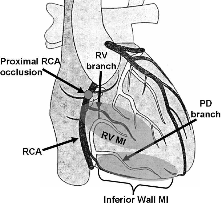
- The right coronary artery (RCA) supplies blood to the inferior wall of the left ventricle via its posterior descending (PD) branch. The right coronary artery also supplies blood to the right ventricular (RV) wall via its RV branch located a short distance from the arteries' origin in the aorta.
右冠状动脉(RCA)通过其后降(PD)分支向左心室下壁提供血液。右冠状动脉还通过其 RV 分支向右心室(RV)壁提供血液,该分支距离主动脉起源不远。
# Assessing RV Function
| Normal | |
|---|---|
Tricuspid Annular Plane Systolic Excursion (TAPSE) | ≥ 16mm |
≥ 35% | |
RV S' (TDI at RV free wall) | ≥ 12cm/s |
Tei index | < 0.4 |
# The Right Atrium
- Assessed in apical four chamber view
心尖四腔切面评估 - Planimetry most accurate way to assess RA (area in cm2)
测面法,评估 RA 的最准确方法(面积以 cm2 为单位)
| 严重程度分类 | |
|---|---|
| Mild | ≥ 18cm2 |
| Moderate | ≥ 28cm2 |
| Severe | ≥ 38cm2 |
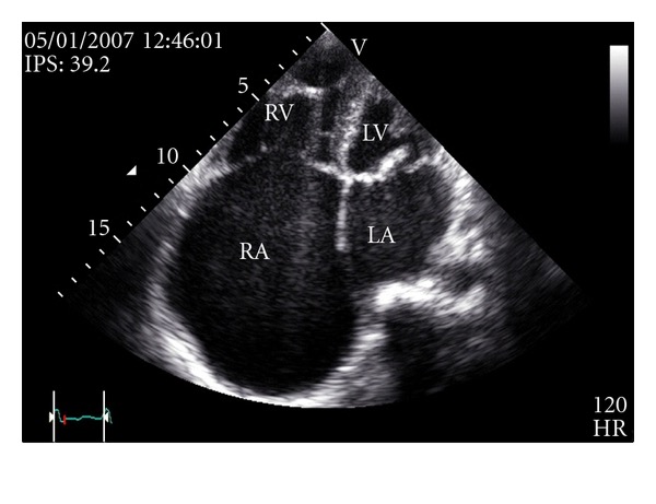
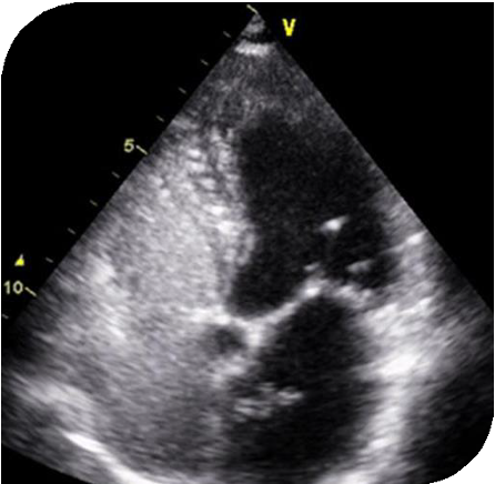
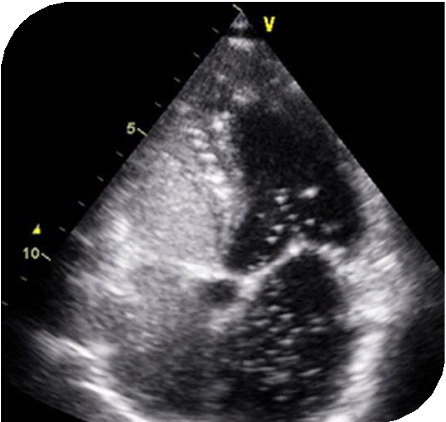
Transthoracic echocardiography, apical four chambers view. Massive enlargement of the right atrium. The right atrial area was 80.6 cm2 (53.7 cm2/m2), and the calculated right atrial volume was 621 mL (414 mL/m2).
经胸超声心动图心尖四腔切面。右心房严重扩大。Causes of RA Dilatation RA 扩张的原因
- Pulmonary Hypertension 肺动脉高压
- Tricuspid valve disease 三尖瓣病变
- Ebstein's anomaly
- Atrial septal defect 房间隔缺损
- Atrial fibrillation 心房颤动
- Dilated cardiomyopathy 扩张型心肌病
# Q&A
EI 测量的位置
偏心指数的测量位置,选用胸骨旁短轴 - 乳头肌水平,也就是左室中段的水平来进行测量
大量或极大量的三尖瓣反流可否用于评估肺动脉高压?
此时用超声心动图来评估肺动脉压力是不准确的。三尖瓣反流频谱呈现典型的短的三角形状的频谱图,因为右房和右室的压差在很短的时间内就达到了平衡;此时用这个图来评估右房压是不准确的