# Learning Objectives
- Basics of Ultrasound
超声波基础知识 - M-mode
- 2D imaging
- Continuous doppler
- Pulse wave doppler
- Colour flow doppler
- Tissue doppler imaging
- Artefact
伪像 / 伪影 - Bubble, contrast and stress echo.
震荡生理盐水,超声增强剂成像和负荷超声心动图
# Properties of Ultrasound Waves 超声波的特性
- Frequency 频率
- Number of cycles per unit time
单位时间内的循环次数
频率与穿透力、分辨率有关 - Wavelength 波长
- Length of a single cycle in mm;
单个周期的长度,单位为毫米;
wavelength = velocity / frequency - Propagation speed 传播速度
- Speed of ultrasound through tissue
超声波通过组织的速度 - Period 时间周期
- Time required for one cycle in microseconds
一个周期所需的时间 (以微秒为单位) - Power 动率
- Rate of energy transfer determined by the ultrasound source - decreases as the beam is propagated.
由超声源决定的能量传递速率 -- 随着波束的传播而降低。
# Wavelength and Frequency 波长和频率
- Wavelength and frequency are inversely related
波长和频率负相关
频率越高,分辨率越好,波长越短,穿透力越差 - Audible sound: 20 kHz
人耳可听到的声音 - Transthoracic echo: 2 - 3 MHz
经胸超声心动图 - Transoesophageal echo: 3.5 - 7 MHz
经食管超声心动图 - IVUS: 10 - 40 MHz.
血管内超声
High frequency ultrasound has better resolution but reduced penetration due to increased scattering.
高频超声具有更好的分辨率,但由于散射增加,穿透力降低。
Low frequency ultrasound has increased penetration but reduced resolution i.e. it is a trade off.
低频超声波增加了穿透力,但降低了分辨率,即这是一种权衡。
如果临床上遇到较瘦的患者,可以适当提高频率,因为不需要很高的穿透力,但可使分辨率变好。
在临床上根据不同病人的体型体态来调节频率,使穿透力和分辨率达到平衡。
# Velocity or Propagation Speed 传播速度
- Propagation speed of ultrasound depends on the density and stiffness/ compressibility of the media.
超声波的传播速度取决于介质的密度和硬度 / 可压缩性。- Air - 330m/s; 空气的传播速度
- Water - 1570m/s; 水的传播速度
- Tissue - 1540m/s; 组织的传播速度(水和组织类似)
- Bone - 4080m/s。 骨的传播速度
Given that the velocity is fixed at 1540m/s we can calculate the wavelength of cardiac ultrasound.
假设速度固定在 1540m/s,我们可以计算心脏超声的波长。
也就是说用超声进行检查时,分辨率不会超过 0.5mm。
如果组织过于细小,小于这个值,在超声上有可能看不清晰或看不到。
# Amplitude and Attenuation 振幅与衰减
Amplitude is the strength of the ultrasound beam (decibels)
振幅是超声波的强度 (分贝)Amplitude decreases by 1dB per 1MHz per 1cm travelled
超声波每传播 1cm,每 1MHz 会减少 1 分贝的振幅Attenuation is frequency dependant - lower frequency ultrasound can penetrate deeper.
衰减与频率有关 - 频率越低,穿透越深(低频率的超声波具有较高的穿透性)
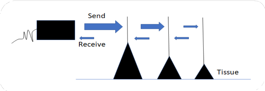
# Piezoelectric effect 压电效应
- Electric energy causes vibration of the piezoelectric crystals within the transducer which creates ultrasound waves.
高频电信号使换能器内的压电晶体振动,从而产生高频的声信号(超声波)。 - Waves are sent from the transducer and reflected off structures in the heart.
超声波从换能器发出,并在心脏的不同结构上被反射回探头。
- Piezoelectric effect 压电效应
- Send waves - Convert electricity to ultrasound waves
发送波 - 将电转换为超声波 - Receive waves - Convert ultrasound waves into electricity
回收波 - 将超声波转换成电能 - The crystals act as a ‘speaker’ and ‘microphone’.
晶体充当 “扬声器” 和 “麦克风”。
- Send waves - Convert electricity to ultrasound waves
# Pulse Repetition Frequency 脉冲重复频率
- Waves are generated in pulses.
超声波以脉冲的形式产生。 - The number of pulses generated per unit time is the pulse repetition frequency
PRF.
脉冲重复频率PRF是指单位时间内产生的脉冲数。 - The PRF must allow for the signal to reach the target and be reflected before the next pulse is generated. This is the ‘listening time’.
PRF 必须满足信号到达目标结构,并被反射接受成功之后,再次发送信号的这一条件。这是 “聆听时间”。
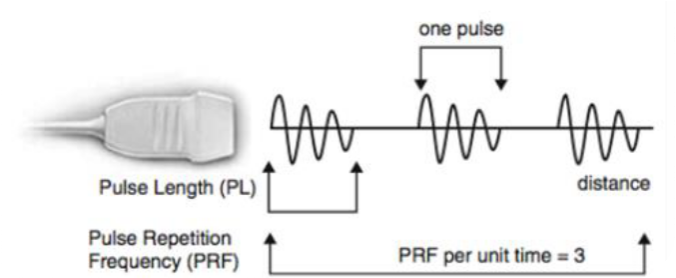
Pulse Length PL 脉冲长度
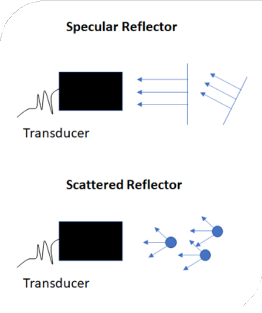
# Reflection 反射
- Specular reflection 镜面反射
- Produced by smooth reflectors which are large compared to wavelength of the US (e.g. cardiac tissue) – greater proportion of sound reflected
由与超声波长相比更大的光滑的反射物 (如心脏、肝脏组织) 产生,- 大部分超声波会被反射回去 - Scattered reflection 散射
- produced by small reflectors or a rough interface (e.g. red blood cells) – more scattering hence reduced proportion of reflection
由较小的反射物或粗糙的表面 (如红细胞) 产生 - 更多的散射,导致成像的反射更弱
# Acoustic Impedance 声阻抗
The amount of ultrasound waves reflected depends on the acoustic impedance of the two media.
超声波的反射量取决于两种介质的声阻抗。Without gel, the air-tissue interface results in >99% of ultrasound waves being reflected and, hence, no image.
在没有凝胶的情况下,空气 - 组织界面导致 99% 以上的超声波被反射而损失掉,因此会不成像。Ultrasound gel has a similar acoustic impendence to tissue and therefore more is absorbed and can image deeper structures.
超声波凝胶的声阻抗与人体组织类似,因此大量的超声波可以被吸收到,并在组织中传播,可以成像更深的结构。
# Transthoracic Echocardiography 经胸超声心动图
- The standard transthoracic echocardiogram window are parasternal, apical, subcostal and suprasternal.
经胸超声心动图的标准声窗包括胸骨旁、心尖、剑突下和胸骨上窝。 - Orientation of the planes in transthoracic echocardiography are long axis, short axis, four, five, two and three chamber.
经胸超声心动图的定位方向有长轴、短轴、四腔、五腔、二腔、三腔。
# Imaging Modes 成像模式
- M-Mode
- 2D imaging
- Continuous wave doppler
- Pulse wave doppler
- Tissue doppler imaging.
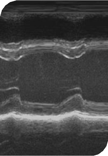
# 1D imaging – M mode
- Single crystal rapidly alternates between sending and receiving modes with rapid updating (>1000Hz)
单晶在发送和接收模式之间快速切换,更新迅速 (>1000 赫兹) - Single scan line 单一扫描线
- High sampling rate – therefore high temporal resolution and can record rapid movements.
有较高的取样率 - 因此具有高时间分辨率,可以记录快速运动。
也可以进行一些径线测量,配合同步的心电图辨别时收缩期还是舒张期
# Uses of M-Mode
临床应用不是很多,但仍然可以用于径线测量
Parasternal long axis 胸骨旁长轴
- Aortic root size and left atrium diameter
主动脉根部大小和左房内径 - Opening of mitral valve leaflets
二尖瓣瓣叶的开放,评估有没有狭窄 - Left and right ventricle dimensions.
左心室和右心室大小
- Aortic root size and left atrium diameter
Apical four chamber 心尖四腔心
- Tricuspid annular plane systolic excursion
TAPSE三尖瓣瓣环平面收缩期偏移,评估右室收缩功能 - Mitral annular plane systolic excursion
MAPSE二尖瓣瓣环平面收缩期偏移
- Tricuspid annular plane systolic excursion
Visualise a rapidly moving structure e.g. flail, mass or vegetation.
可视化快速移动的结构,例如连枷、块或赘生物。
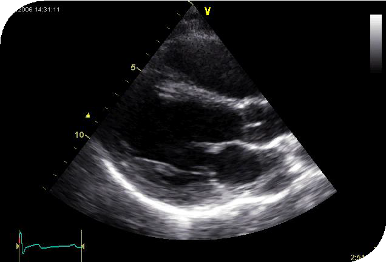
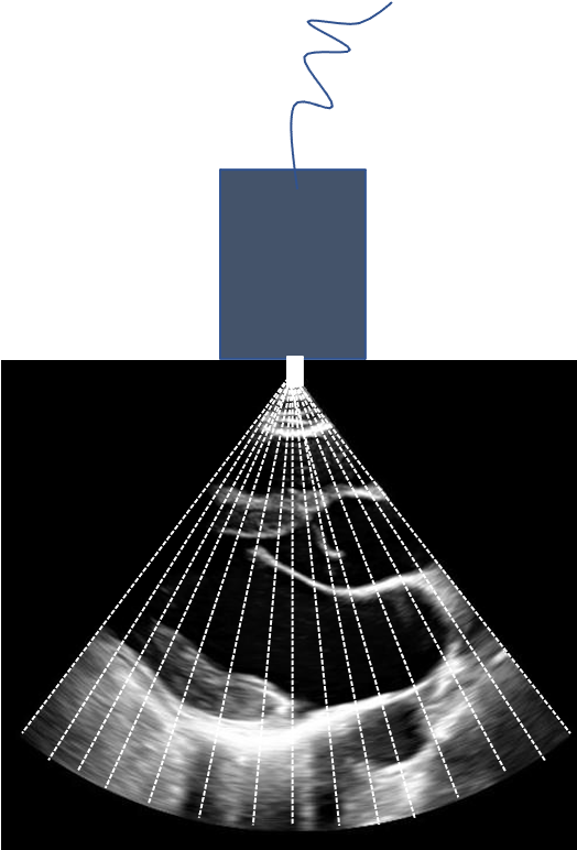
# 2D imaging 二维成像
US pulses are sent down a series of scan lines from an array of small piezoelectric crystals
超声脉冲从小型压电晶体阵列沿一系列扫描线向下发送Waves are reflected back to the same array generating a 2D image of the region being scanned
超声波被反射回同一压电晶体阵列,生成被扫描区域的 2D 图像High intensity signals - White
高强度信号 - 白色Medium - Grey
中等强度信号 - 灰色Low intensity - Black.
低强度信号 - 黑色
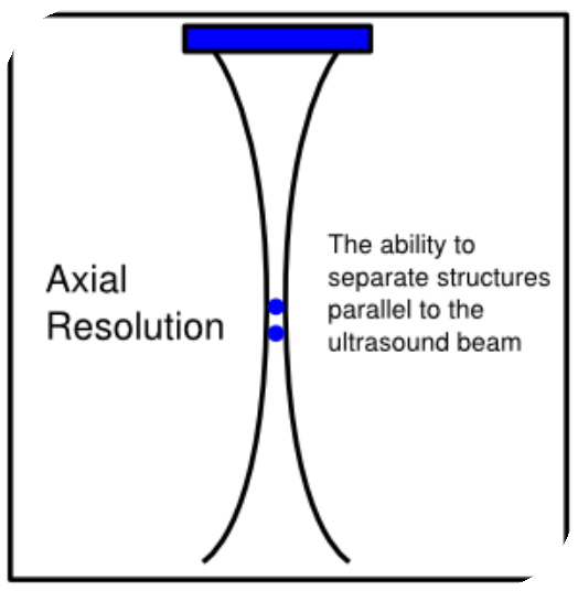
# Spatial Resolution - Axial 空间分辨率 - 轴向
- Axial resolution 轴向分辨率
- The ability to separate structures parallel to the ultrasound beam
区分平行于声波束的组织结构的能力
Smallest distance between two points that allows the objects to be distinguished as separate along the line of the beam
允许沿光束线将对象独立区分的两点之间的最小距离Higher frequency and shorter pulse length = increased axial resolution
更高的频率和更短的脉冲长度 = 这两个点越接近 = 提高轴向分辨率Axial resolution is highest at the beam focus.
轴向分辨率在光束聚焦处最高。
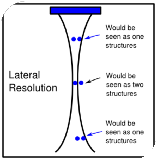
# Spatial Resolution - Lateral 空间分辨率 - 侧向
- Lateral resolution 侧向分辨率
- Ability to distinguish two adjacent objects that are perpendicular to the beam
区分垂直于光束的两个相邻物体的能力
Beam width, depth and gain affect lateral resolution
光束宽度、深度和增益影响侧向分辨率Lateral resolution is highest at the beam focus.
侧向分辨率在光束聚焦处最高。
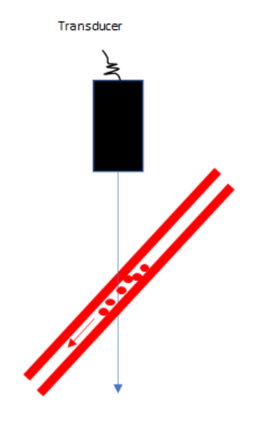
# “Drop Out” 缺失
“Drop out” occurs when the US beam is parallel to the structure being imaged
当超声束与成像的结构平行时,会发生 “脱落”。
In the subcostal window the ultrasound beam is perpendicular to the septum and therefore the interatrial septum is clearer.
在剑突下声窗中,超声束垂直于房间隔,因此房间隔更清晰。


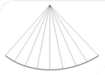
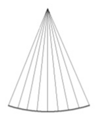
# Temporal Resolution 时间分辨率
Ability to track moving targets over time
追踪移动结构随时间运动的状况的能力Dependant on having high frame rate
取决于高帧率;帧频越高,时间分辨率越高Increased by: 提高时间分辨率通过:
- Reducing depth 降低成像深度
- Reducing sector width and maintaining scan line density. 降低扇区宽度并保持扫描线密度。
# Contrast Resolution 对比分辨率
- Gain 总增益
- Affects brightness of image 影响图像亮度
- Increases noise and therefore reduced resolution. 增加噪音,从而降低分辨率。
- Time Gain Compensation 时间增益补偿
- Deeper structures are poorly visualised due to attenuation
由于衰减的缘故,组织结构越深越不容易被观察到 - TGC can be used to provide compensatory amplification of the weaker signals coming from the deeper tissues to achieve a uniform image.
TGC 可以用来补偿,对深层组织的微弱信号进行放大,以获得均匀的图像。
- Deeper structures are poorly visualised due to attenuation


# Doppler Echocardiography 多普勒超声心动图
- Determine speed and direction of movement of red blood cells or tissues
可以用来测定红细胞或组织的运动速度和方向 - Doppler signal should be parallel with the flow of blood
多普勒信号应与血流方向尽量平行 - Higher doppler frequency is obtained if:
如果满足以下条件,则可获得更高的多普勒频率:- Higher blood velocity
更快的血流速度 - Beam is aligned to flow direction
波束与流动方向对齐 - Higher beam frequency.
更高的波束频率。
- Higher blood velocity
# Aliasing 混叠
- Ultrasound system is trying to image an event that is occurring faster than the rate of sampling
当成像目标的移动速度大于超声波系统的取样速度时,会产生混叠现象 - Velocity exceeds the Nyquist Limit
当速度超过尼奎斯特极限时 - Image displayed suggests flow is heading in the opposite direction
成像表明血流正朝着相反的方向运动 - Seen in both colour and pulse wave doppler.
彩色多普勒和脉冲波多普勒中都可以看到
如果血流的方向是朝向探头,应该表现为红色,如果血流方向是远离探头的,则表现为蓝色。
当血流速度超过尼奎斯特极限时,就会表现为反方向。
比如瓣膜存在狭窄或者反流的情况下,当血流速度比较高时,就可以出现这种混叠的现象。

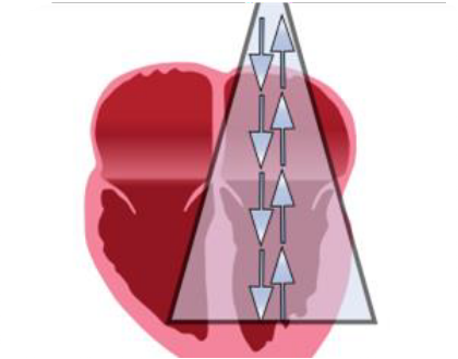
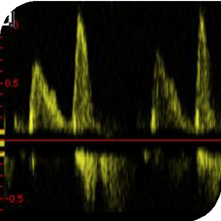
# Continuous Wave Doppler 连续波多普勒
- Crystal continuously transmits and receives ultrasound signals
是晶体连续发射和回收信号 - High velocities can be accurately measured
可以精确测量高速度血流 - No selectivity or depth discrimination i.e. poor range resolution
没有选择性或深度分辨率,即范围分辨率较差,无深度位置区分 - Can assess large sample volumes
可以评估大的取样容积,会捕捉整个取样线上的速度 - Less prone to aliasing than pulse wave doppler.
与脉冲波多普勒相比,不易出现混叠。
当对主动脉瓣狭窄测量经主动脉瓣的最大血流速度时,就运用了连续多普勒来获得最大的血流速度
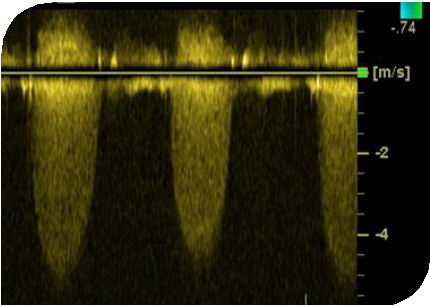
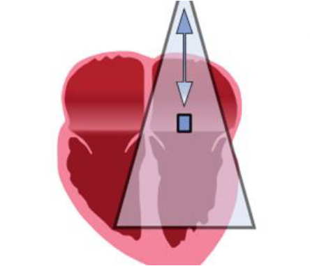
# Pulse Wave Doppler 脉冲多普勒
- A crystal sends a signal and the same crystal waits to receive the signal
晶体发送信号,同一晶体等待接收信号 - Good range resolution - signals from a specific distance can be selected
良好的范围分辨率 - 可以选择特定距离的信号进行取样 - Prone to aliasing and therefore cannot be used to assess high velocity flow.
容易产生混叠,因此不能用于评估速度较高的血流速度。
# Colour Doppler 彩色多普勒
- A form of pulse wave doppler 脉冲波多普勒的一种形式
- Same advantages and limitations as PW doppler:
与 PW 相同的优点和局限性:- Good range resolution
良好的范围分辨率 - Aliasing if high velocity
如果速度较高,则会产生混叠
- Good range resolution
- Visual assessment of valve disease and shunts.
瓣膜疾病和分流的视觉评估。


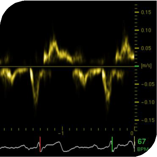
# Tissue Doppler Imaging 组织多普勒成像
- Tissue doppler targets myocardial tissue
组织多普勒的目标是心肌组织 - Picks up low velocity and high signal amplitude (therefore does not measure blood flow)
拾取低速度和高振幅信号 (因此不测量血流量) - Can be used for assessment of diastolic function and RV function.
可用于评价舒张功能和右室功能。
# Artefact 伪像 / 伪影
在成像时需要区分哪些是真实存在的,哪些是由于成像的一些局限性而造成的一个伪像。
伪像在临床中非常见,比如:
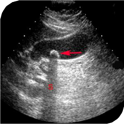
- Acoustic shadow 声影
- Signal void behind structures that strongly absorb or reflect ultrasound waves e.g. calcified valve.
强烈吸收或反射超声波的结构的后面的信号空洞,例如钙化的瓣膜。
当存在钙化时,信号会被钙化组织强烈地吸收反射,在它的远端或是下方,就可以看到一个阴暗的区域,如同影子一般,称作为声影。![]()
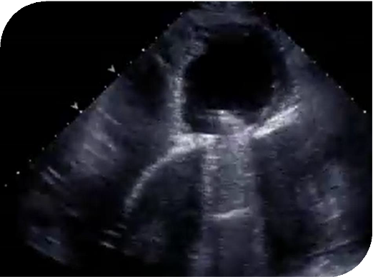
- Reverberation 混响
- Ultrasound beam reflects back and forth between two parallel reflectors (reverberates).
超声波束在两个平行反射器之间来回反射(混响)。
当超声信号在这两个平行的墙的反射结构中来回进行反射,就会形成两个类似于镜子一样的成像,称为混响。
Waves are delayed in returning and interpreted as a deeper structure.
超声波返回延迟,并被解释为更深的结构。![]()
# Advanced imaging techniques 附加技术
# Bubble Echocardiography 微泡超声心动图
- Agitated saline injected peripherally
外周静脉注射震荡生理盐水(7ml 生理盐水,1ml 空气,以及部分患者的血液进行混合,形成微泡) - Bubble diameter 50-90µm
微泡直径 50-90µm - Destroyed as they pass through pulmonary capillaries (unless there is a pulmonary shunt)
微泡在通过肺毛细血管时被破坏清除(除非有肺分流) - Can diagnose PFO, ASD, persistent left superior vena cava.
可诊断卵圆孔未闭、房间隔缺损、永存左上腔静脉。
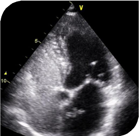
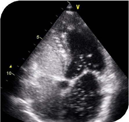
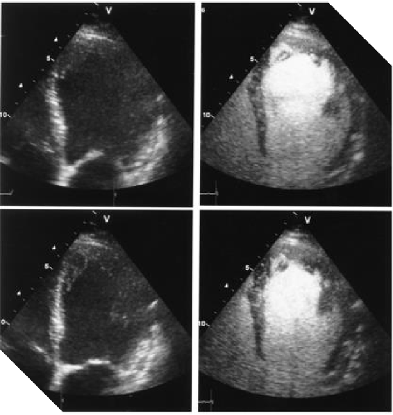
# Contrast Echocardiography 超声增强剂成像
如果成像质量不好,无法观察左室运动的真实情况
- Diameter of contrast microbubble is 1-5µm
增强剂(声诺维)微泡直径 1-5µm - Can pass through pulmonary circulation to the LV
可以通过肺循环进入左室,能更高的观察左室心内膜的情况 - Improves accuracy of LV volume and ejection fraction(EF) assessment.
提高左室容积和射血分数评估的准确性
# Stress Echocardiography 负荷超声心动图
- Dobutamine or exercise stress
包括药物性(多巴酚丁胺)负荷或运动负荷 - Can evaluate inducible ischaemia
可以评估诱导性心肌缺血 - Assess true severity of low flow low gradient aortic stenosis
评估低流量低跨瓣压主动脉瓣狭窄的真实严重程度 - Assess viability of myocardium.
评估心肌的生存能力