# Echo Basics and the Right Ventricle
With regard to the anatomy of the RV, which of the following statements are true?
关于 RV 的解剖结构,以下哪项陈述是正确的?
- Wall thickness is the same as the LV 室壁厚度与 LV 相同
- In the apical four chamber view, the LV appears more trabeculated 在心尖四腔切面,左心室似乎由更多肌小梁
- The RV is anterior to the LV 右室在左室的前面
- The RV is supplied by the left circumflex artery 右室由左旋支动脉供血
- The structure of the RV is symmetrical 右室的结构是对称的
左室室壁比右室厚;
右室肌小梁更多
右室为右冠为主,及左前降支供心尖部的血液
右室是非对称的,新月形形状
Which of the following are causes of pulmonary hypertension?
以下哪项是肺动脉高压的原因?
- Mitral valve disease 二尖瓣疾病
- Diastolic dysfunction 舒张功能障碍
- Systolic dysfunction 收缩功能障碍
- Left to right shunts 左向右分流
- Pulmonary embolism 肺栓塞
左心压力高、舒张功能、收缩功能障碍都可以导致肺动脉高压
Which of the following are true?
- TAPSE > 14mm is considered normal
- RV base measurement > 4.4cm is normal 右室基底部测量
- RV diastolic area of 28cm2 is abnormal 右室舒张末面积
- Fractional area change of 32% is normal 面积分数
- PA diameter of 1.8cm in the parasternal short axis is considered normal 胸骨旁短轴切面的肺动脉内径
TAPSE > 16mm 认为正常
RV base measurement < 4.2cm 认为正常
RV diastolic area 28 cm2 认为有轻度扩张,一般上限是 25 cm2
FAC 一般要求 35% 以上算正常,有的指南认为,男性一般大于 30%,女性大于 35%
PA 直径 < 2.3cm 认为是正常
You are asked to perform an echocardiogram on a 25-year-old male who is breathless. His TR Vmax is 4m/s. His IVC is 1.2cm with complete collapse on inspiration. What is his PASP?
为一名 25 岁的气急男性做超声心动图检查。TR Vmax 为 4m/s。IVC 为 1.2 cm,呼气末完全塌陷。他的 PASP 是多少?
- 34 – 39mmHg
- 54 – 59mmHg
- 44 – 49mmHg
- 64 – 69mmHg
- 24 – 29mmHg
估算方法
4×4×4+(0 5)
You perform an echocardiogram on a 62-year-old man. You find moderate pulmonary regurgitation with a peak velocity of 4.2m/s and end-diastolic peak velocity of 3m/s. The IVC is dilated at 2.6cm and collapses < 50%. Which of these statements are true?
为一名 62 岁的男子做超声心动图检查。发现中度肺动脉瓣反流,峰值速度为 4.2 m/s,舒张期末峰值速度为 3 m/s。IVC 扩张到 2.6 cm,呼吸塌陷率 < 50%。以下哪项陈述是正确的?
- You can calculate the PASP from the information above 可以根据以上信息计算 PASP
- The diastolic PA pressure is 36 + RAP
- The diastolic PA pressure is 64 + RAP
- The mean PA pressure is 64 + RAP
- The mean PA pressure is 36 + RAP
所以要算 通过 PR jet 可以计算 dPAP 肺动脉舒张压 或 mPAP 平均肺动脉压
dPAP=PR end diastolic pressure gradient+RAP=4×3×3+RAP=36
mPAP=PR peak pressure gradient+RAP=4×4.2×4.2+RAP=70.56
Which of the following may be found in severe pulmonary hypertension?
严重肺动脉高压患者可能会出现以下哪种情况?
- TR Vmax of 4m/s
- Mid-systolic closure of pulmonary valve 肺动脉瓣收缩中期关闭
- Mid-systolic notch on PA doppler 肺动脉 PW 收缩中期呈现刻痕状改变
- A fixed and dilated IVC 固定并扩张的下腔静脉
- Dilated right ventricle 右心室扩张
Which of the following are true?
- Tissue doppler can be used to calculate myocardial performance index of the right ventricle 组织多普勒可用于计算右室心肌功能指数
- McConnell’s sign may be seen in acute PE 急性肺栓塞可出现麦康奈尔征
- Localised aneurysms of the RV may be seen in AVC 右室局限性室壁瘤可见于致心律失常性心肌病
- Arrythmogenic cardiomyopathy (AVC) only occurs in the RV 致心律失常性心肌病(AVC)仅发生于 RV
- A Tei index of 0.6 indicates normal RV function Tei 指数 0.6 表示 RV 功能正常
致心律失常性心肌病也可能发生在左室
使用组织多普勒测 Tei 指数一般的界限值是 0.55,使用 PW 测一般是 0.4
Which of the following are true?
- Stroke volume of the RV is around 10% less than that of the LV in a normal heart 正常心脏的右室每搏量比左室少 10% 左右
- In Ebstein’s anomaly, the RV is small and the RA is larger than usual 在 Ebstein 畸形中,RV 较小,RA 较大
- ARVC causes fatty infiltration of the right ventricle ARVC 导致右心室脂肪浸润
- An RV EF of 45% is normal 右室射血分数为 45% 是正常的
- The area-length method for EF calculation is accurate in RV assessment 面积 - 长度法计算的 EF 在 RV 评估中是准确的
根据连续方程式,进入左室的血液等于右室,也就是左右室的每搏量是相等的
左右室 EF 值的评估方法是不一样的,因此参考值有差异;
且右室评估 EF 时,因为其形态问题,没有把所有的射血量都算进去,所以 EF 的评估也不是准确的
Which of the following are true?
- Eccentricity index of >1 in systole indicates pressure overload 收缩期偏心指数 > 1 表示压力超负荷
- Eccentricity index of >1 in diastole indicates volume overload 舒张期偏心指数 > 1 提示容量超负荷
- Eccentricity index > 1 in both diastole and systole indicates pressure overload 舒张期和收缩期偏心指数均 > 1 表示压力超负荷
- A dilated RA is a prognostic feature of severe PH 右房扩张是重度 PH 的预后特征
- A pericardial effusion is a prognostic feature of severe PH 心包积液是重度 PH 的预后特征
An 89-year-old man collapses in the street following a long haul flight. You perform an echocardiogram and find the base of the RV is 4.4cm, TAPSE 1.2cm, RV S’ 10cm/s, TR Vmax 4.5m/s.
一名 89 岁的老人在长途飞行后晕倒在街上。做超声心动图,发现右室基底部 4.4cm, TAPSE 1.2 cm,RV S‘10 cm/s,TR Vmax 4.5m/s。
Which of the following are true?
- His RV systolic function is normal 右室收缩功能正常
- His RV is severely dilated 右室严重扩张
- His RV free wall might be akinetic 右室游离壁可能无运动
- His RV apex might be akinetic 右室心尖部可能无运动
- He has mildly raised pulmonary artery systolic pressures 他有轻微的肺动脉高压
TAPSE 降低,虽然组织多普勒基本正常,但可以判断右室收缩功能是异常的,但没有严重扩张,基底部 4.4cm 是一个临界扩张
TAPSE 测的是三尖瓣,右室的游离壁有可能出现运动正常,心尖部倒有可能出现无运动
伯努利方程计算可以得到重度肺动脉高压的表现
Which of the following are true?
- The right atrium does not dilate in pulmonary hypertension 肺动脉高压时右心房不扩张
- Right atrial area of 17cm2 is considered normal 右心房面积 17cm2 视为正常
- Right atrial area of 38cm2 is considered severely dilated 右心房面积 38cm2 视为严重扩张
- Right atrial pressure can be assessed based on IVC diameter and collapse on inspiration 可以根据 IVC 直径和吸气塌陷回缩率来评估右心房压力
- Raised right atrial pressure may be due to pulmonary hypertension 右心房压力升高可能是由于肺动脉高压
Which of the following statements are true?
- Frequency = wavelength/velocity 频率 = 波长 / 速度
- The frequency of transthoracic echo is 2-3MHz 经胸超声频率为 2~3 MHz
- Increasing the frequency results in better penetration of ultrasound waves 提高频率可提高超声波的穿透性
- The velocity of ultrasound in cardiac tissue is 1570m/s 超声在心肌组织中的速度为 1570 m/s
- Attenuation is dependant on the frequency of ultrasound 衰减取决于超声波的频率
频率 = 速度 / 波长
增加频率时分辨率是增加的,但是穿透力是下降的。
超声在心肌组织中的传播速度是 1540 m/s
Which of the following statements are true?
- Specular reflection is produced by reflectors which are larger than the wavelength 反射是由大于波长的反射器产生的
- Scattered reflection is produced by reflectors which are smaller than the wavelength 散射是由小于波长的反射器产生的
- M-mode has a low sampling rate M 模式的采样率很低
- Piezo-electric crystals are not used in modern ultrasound probes 压电晶体不用于现代超声波探针
- High intensity signals appear black on ultrasound 高强度信号在超声波上显示为黑色
M 模式其实有很高的采样率,所以它的时间分辨率是很高的。
Which of the following statements are true?
- Axial resolution is the ability to separate two adjacent structures which are perpendicular to the beam 轴向分辨率是分辨和超声束相垂直的两个点的能力
- Lateral resolution is the ability to separate structures parallel to the ultrasound beam 横向分辨率是分辨与超声束相平行的结构的能力
- Axial resolution is increased at higher frequencies 在较高频率下,轴向分辨率增加
- Temporal resolution is the ability to track moving objects over time 时间分辨率是随着时间推移跟踪移动对象的能力
- Temporal resolution increases with a lower frame rate 帧频越低,时间分辨率越高
轴向分辨率是分辨和超声束相平行的两个点的能力,超声表现为上下两点
横向分辨率是分辨和超声束相垂直的结构的能力,在超声上表现为左右两点
帧速率越高,时间分辨率越高,所以可以减少感兴趣扇区的面积深度、宽度,这样可以提高帧频,从而提高时间分辨率
Which of the following statements are true?
- Aliasing occurs because the system is try to image an event occurring faster than the rate of sampling 产生混叠的原因是系统尝试以比采样速率更快的速度对发生的事件进行成像
- Aliasing affects continuous wave doppler more than pulse wave doppler 混叠对连续波多谱勒的影响大于脉冲波多谱勒
- Doppler echocardiography only gives information on speed of movement 多普勒超声心动图仅提供有关运动速度的信息
- Doppler signal should be perpendicular to the flow of blood to yield maximum velocity 多普勒信号应垂直于血流以产生最大速度
- Higher doppler frequency is obtained if there is lower blood velocity 血流速度越低,多谱勒频率越高
混叠影响脉冲多普勒多于连续多普勒。
多普勒超声心动图不但提供速度的信息,还提供方向的信息。
在做连续多普勒时,尽量要平行于血流的方向,因为它有角度依赖问题,这样才可以取得尽量高的速度。
较低的血流速度,一般取得的是较低的多普勒频谱。
# Clinical Case: The Right Ventricle
48-year-old male
Background: Diabetes mellitus
Medications: Metformin
48 岁的男性有糖尿病史,使用二甲双胍
Presents with episode of collapse
Normal chest radiograph
ECG shows non-specific ST changes
Troponin is 56 (normal 0-40)
数次出现晕倒。胸部 X 片表现为正常,心电图无特异性的 ST 段改变,肌钙蛋白的测量值是 56。
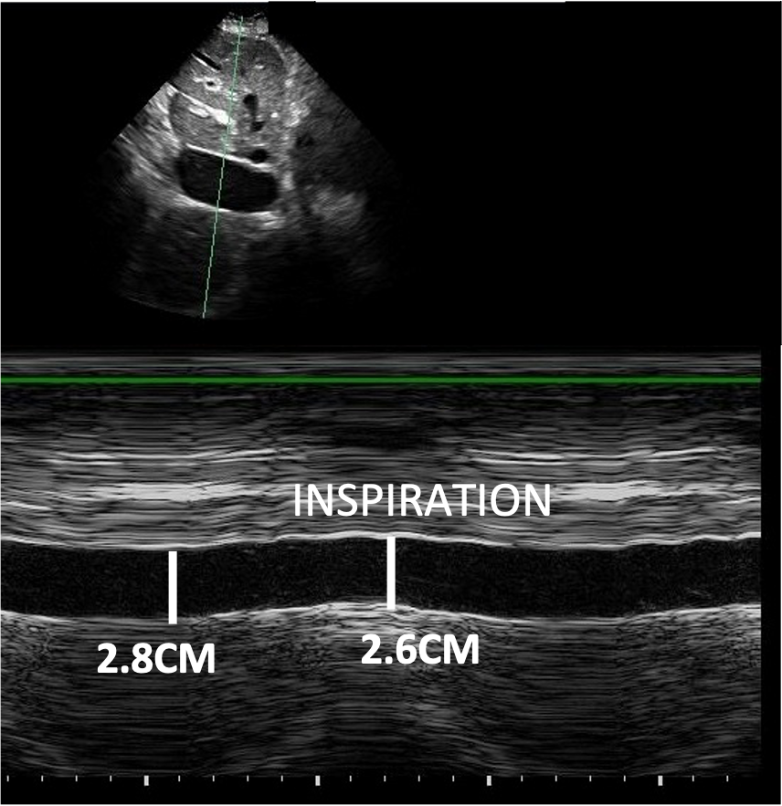
You are asked to perform an echocardiogram


![]()

![]()
The right atrial pressure is:
- 0-5 mmHg
- 5-10 mmHg
- 15-20 mmHg
- 10-15 mmHg
IVC 存在扩张,回缩率远小于 50%。
The right ventricle is:
- Mildly dilated
- Moderately dilated
- Severely dilated
- Normal in dimensions
图 2 右室基底部内径 = 4.4cm
Visually, the RV systolic function is:
- Mildly impaired
- Normal
- Severely impaired
- Unable to assess
here visually it may well be normal in longitudinally anyway
目测是正常的,还需要测量 TAPSE、FAC 等
By colour doppler, the patient has:
- Mild TR
- Severe TR
- Moderate TR
- No evidence of TR
Very significant leak
The TR max pressure gradient is:
- 28 mmHg
- 25 mmHg
- 30 mmHg
- 34 mmHg
- 27 mmHg
根据 the simplified benulee equation, TR Vmax = 2.6m/s,可以计算出跨瓣压差的压差是 27.04mmHg。
The pulmonary artery systolic pressure is:
- 33 – 38 mmHg
- 42 – 47 mmHg
- 56 – 61 mmHg
- 49 – 54 mmHg
在 27 的基础上加 15 到 20 mmHg 的右房压,就可以得到这个 42 到 47 mmHg 的肺动脉收缩压。
The patient has evidence of:
- RV volume overload
- RV pressure overload
- A normal RV
- Left ventricular systolic impairment
偏心指数在收缩期 > 1 提示右室压力负荷过高。
The likely diagnosis is:
- Atrial septal defect
- Pulmonary embolism
- ARVC
- Ebstein’s anomaly
- Left ventricular systolic impairment
根据病史和超声表现
# Echo Basics Part 2
The following LV segments can be seen in the Long Axis View:
在胸骨旁长轴切面可以看到以下哪些左室节段:
- Basal Inferolateral 下侧壁基底段(下侧壁 = 后壁)
- Apical Anterior 前壁心尖段
- Mid Anteroseptal 前间隔中段
- Apical Anteroseptal 前间隔心尖段
- Mid Inferior 下壁中段
首先胸骨旁长轴的中间是前间隔,它的下方是下侧壁,可以看到下侧壁的基底段和中段。
心尖段在胸骨旁长轴是看不到的
能看到的是下侧壁,也就是后壁,而不是下壁
The following cause inaccuracy in Fractional Shortening Calculation:
以下原因哪些导致了缩短分数计算不准确:
- Irregular Heart Rate 心律不齐
- Slow Heart Rate 心率慢
- Off-axis Images 偏轴成像
- Left Ventricular Hypertrophy 左心室肥厚
- Poor endocardial definition 心内膜清晰度差
为了测量准确,并不乐意看到因为心律不齐导致室壁运动不规律,希望是心律齐 + 心率慢
偏轴成像意味着采样位置不够好
左室肥厚没有什么影响
清晰度差则不能很好的测量计算
Contrast micro bubbles reach the left heart because:
微泡对比剂到达左心是因为:
- They are very large 它们非常大
- They are injected into an artery 它们被注射到动脉中
- They survive passage through the lungs 它们通过肺部幸存下来
- They survive interference by ultrasound 它们在超声波的干扰下幸存下来
- They have the same density as blood 它们的密度和血液一样
The following cause inaccuracy in M-Mode techniques:
以下哪些因素会导致 M - 模式技术不准确:
- Poor penetration of myocardium compared to 2D 与 2D 相比,心肌穿透能力较差
- Irregular heart rate 心律不齐
- Slim patient 纤瘦的患者
- Experienced operator 有经验的操作者
- Artefact from heart valves 心脏瓣膜的伪影
M 模式和 2D 的心肌穿透能力一样
纤瘦的患者和有经验的操作者更容易获得准确数据
心脏瓣膜不足以造成足够的伪影
LV contrast is coated with powder because:
LV 对比剂表面裹以干粉
- Survives passage through pulmonary circulation 通过肺循环存活下来
- Survives destruction by heart valves 从心脏瓣膜的破坏中存活下来
- Survives passage through the left ventricle 从左心室中存活下来
- Becomes more bright 变得更加明亮
- Becomes more dense 变得更加密集
这是唯一的原因
Left ventricular function:
左心室功能包括:
- Circumferential thickening 内向增厚
- Longitudinal lengthening 纵向的伸长
- Longitudinal shortening 纵向的缩短
- Apical thinning 心尖变薄
- Rotation 扭转运动
In Simpson’s rule:
- Disc Height = LV diameter × number of discs
- The discs are shorter towards the apex 向心尖方向的圆盘较短
- LV volume = (IVS + LVIDd + PW)2
- Myocardial volume = Blood pool volume - LV volume
- Individual disk volume = height × disk area 圆盘容积等于高度 × 面积
左室长径除以圆盘数量,不是左室直径
每个圆盘都等高
左室容积的计算应该是立方运算
心肌体积 = 包含心外膜的左室体积 - 血池体积
Left Ventricular Anatomy: 左室解剖
- Inlet, outlet, apical trabecular and basal trabecular portion 流入部、流出部、心尖肌小梁和基底部肌小梁
- The portions have distinct borders 这些部分有明确的界限
- Thickest at the apex 心尖部最厚
- Longitudinal muscle strands in the subepicardium 纵向的肌束分布在心外膜的下方
- Circumferential strands in the middle of the myocardium 环形的肌束在心肌的中层
左室不包括基底部肌小梁
没有明确界限
心尖部最薄
心外膜是斜形的,中层是环形的,心内膜下的是纵向的
LVEDD = 50 mm, LVESD = 40 mm. FS = ?
缩短分数等于舒张末内径减收缩末内径的差值除以舒张末内径。
LVOT radius =1.05 cm LVOT area =?
- 2.5 cm2
- 3.0 cm2
- 3.5 cm2
- 4.0 cm2
- 4.5 cm2
面积公式
LVOT area = 3.5 cm2, TVI = 9.5 cm, Heart rate = 60 bpm (Approximate) Cardiac output = ?
- 1 litre/min
- 1.5 litre/min
- 2 litres/min
- 2.5 litres/min
- 3.0 litres/min
已知左室流出道的面积以及 LVOT 的速度、时间积分,还有心率,计算一下这个心输出量大约是多少。
心输出量等于每搏量乘以心率。每搏量可以根据 LVOT 的面积乘以速度时间积分,所以这三个值相乘,就可以得到心输出量。
Agitated saline contrast is a mixture of:
震荡生理盐水实验用的是哪一种增强剂?
- Saline, 1ml air, 1ml dextrose, 1ml patient’s blood
- Saline, 1ml Sonovue, 1ml patient’s blood
- Saline, 1ml air, 1ml Sonovue
- Saline, 1ml dextrose, 1ml air
- Saline, 1ml air, 1ml patient’s blood
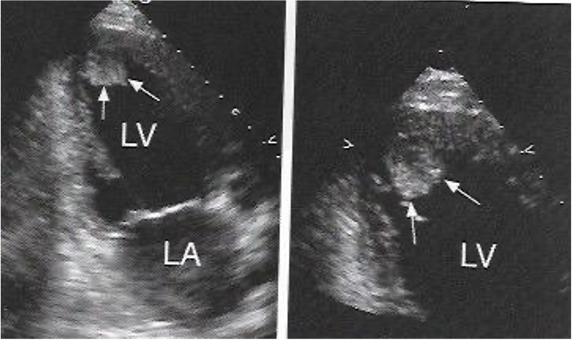
What is the abnormality?
![]()
- Apical hypertrophy 心尖肥厚
- Normal LV 正常左室
- Anterior STEMI 前壁心梗
- LV thrombus 左室血栓
- VSD 室间隔缺损
一般心尖血栓不太容易观察到,可以用增强剂来鉴别
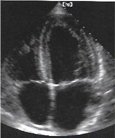
What is the abnormality?
![]()
- Apical HCM
- Normal LV
- Anterior STEMI
- LV thrombus
- VSD
可以用排除法
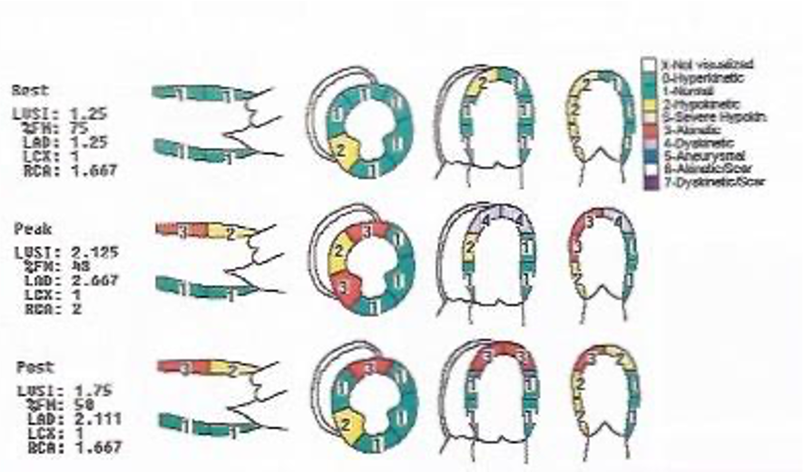
Number of akinetic LV segments at rest
静息状态下无运动的 LV 节段数
![]()
这是多巴酚丁胺负荷实验的截断评分。第一行是静息状态下的评分,第二行是运动负荷在顶峰时的评分,第三行是恢复期的评分。题目问的是在静息状态下的无运动的截断有几个?
red 是 akinetic 的区域,第一行并没有
1 = 正常,绿色
2 = 运动功能降低,黄色,如果严重降低,则橘红色
3 = 无运动,红色
4 = 反向运动
5 = 室壁瘤
6 = 无运动伴疤痕,白色
7 = 反向运动伴疤痕,紫色
# Clinical case
65-year-old male
65 岁的男性
History of MI × 2
Each time treated with stents
心梗两次都是介入放置支架
Breathless on exertion
劳累时呼吸困难
Mild chest tightness but can continue walking
轻度胸闷,但可以继续行走
On dual antiplatelets, statin, beta blocker, nitrate, ACE inhibitor
双抗血小板、他汀类、β 受体阻滞剂、硝酸盐、ACE 抑制剂
SR, HR60 bpm, BP 105/60 mmHg
窦性心律,心率,血压

Most errors in LVEF estimation with:
- FS
- TEI index
- Simpson’s rule
- Mitral valve annular motion towards apex
- Cardiac MRI scan
Least errors in LVEF estimation with:
- FS
- TEI index
- Simpson’s rule
- Mitral valve annular motion towards apex
- Cardiac MRI scan
Assign a wall motion score to the apical IVS:
给心尖部室间隔指定一个室壁运动评分
可以看到左室呈显著扩张,而且呈现球形的外观。
虽然不是动图,但仔细看室间隔以及室间隔的心尖部,可以看到心尖部明显呈现变薄变亮的超声成像,提示有大量的疤痕组织存在。所以答案是给的 E ,6 分的无运动伴疤痕。
LV contrast useful here because:
左室增强剂有用的原因
- The endocardium is not visualised well 心内膜显示不清楚
- The LV is dilated and does not fit in the picture 左心室扩张,与图片不符
- The mitral valve does not close properly, hiding LV 二尖瓣关闭不好,掩盖了部分左心室
- A thick papillary muscle hides part of LV cavity 肥厚的乳头肌隐藏了部分左心室腔
- There may be an apical thrombus present 可能存在心尖血栓
主要要排除一下心室心尖部的血栓,因为左室心尖部呈现疤痕以及无运动的情况。
且心尖部非常扁平的,有可能存在附壁血栓,所以要用增强剂进行一下检查,排除心尖部血栓的存在。