# Evaluation of the Left Ventricle
# Fractional shortening FS 短轴缩短率 / 缩短分数
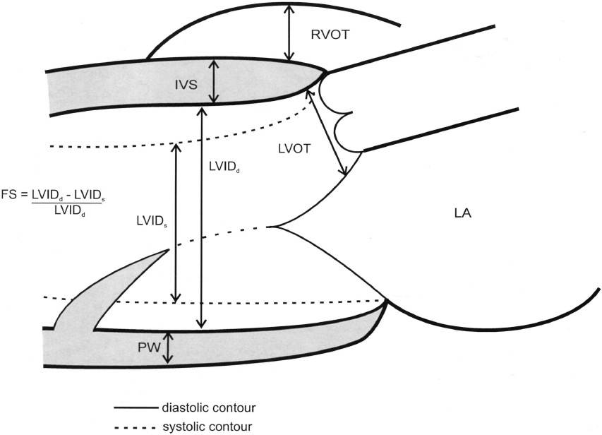
| EDD: end diastolic diameter | 舒张末期内径 |
| ESD: end systolic diameter | 收缩末期内径 |
| EF: ejection fraction | 射血分数 |
| RVOT: right ventricular outflow tract | 右室流出道 |
| IVS: interventricular septum | 室间隔 |
| LVIDd: left ventricular internal diameter at diastole | 左室舒张末期内径 |
| LVIDs: left ventricular internal diameter at systole | 左室收缩末期内径 |
| PW: posterior wall | 后壁 |
| LVOT: left ventricular outflow tract | 左室流出道 |
| LA: left atrium | 左心房 |
- 用于评估左室功能的指标
- Long axis view 选择胸骨旁长轴观,在二尖瓣腱索水平进行评估
- for % 换算成百分比形式,即为缩短分数
- Double for EF. 乘 2 得到 EF
- When measuring ventricular septal thickness, caution is advised to avoid measuring the most proximal portion of septum, which is frequently an area of isolated hypertrophy and angulation that does not truly represent ventricular wall thickness.
测量室间隔厚度时,应注意避免测量室间隔的最近端部分,这通常是一个孤立性肥大和成角的区域,并不真正代表室壁厚度。
# Fractional Area Change FAC 面积变化分数
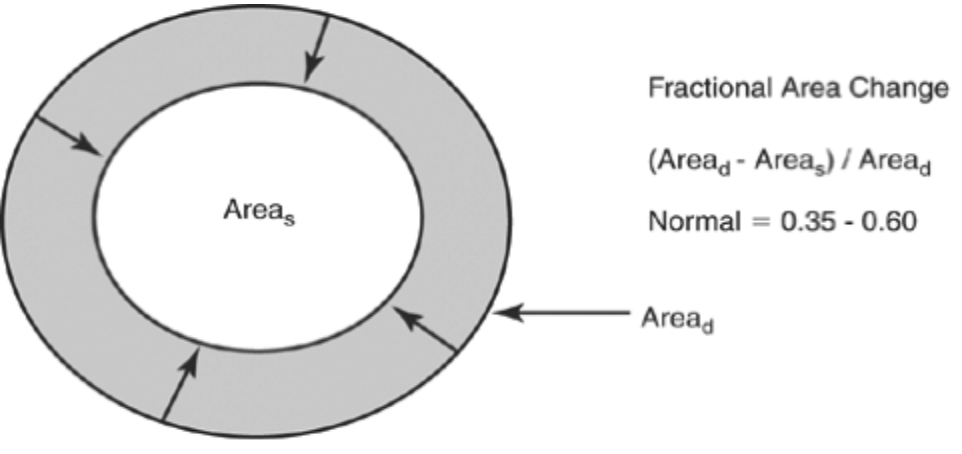
- Used for the Left Ventricle 用于评估左室的收缩功能,也可以用于右室的
- Short axis view 短轴观 (二尖瓣瓣尖水平)
- Compares systolic with diastolic area. 比较收缩压和舒张期面积来计算
- 正常值范围约 35% - 60%
# Simpson’s rule (rule of disks) 辛普森积分法 / 圆盘法则
- Based on the assumption that the chamber will adhere to a predictable shape.
假设左室遵循可预测形状。有的设置为球形,有的设置为圆锥形 - The ventricle is divided into a series of disks of equal height.
将心室分为一系列等高的圆盘。(一般为 10 个)
;- .
。
Equations:
- 圆盘高度 Disc height = h
- 左心室高度 Total LV height = L
- 圆盘数量 Number of discs = n
- 圆盘半径 Disc radius = r
- ;
- 圆盘的容积 ;
- 总容积 .

- In the upper panel, a schematized left ventricular volume has been subdivided into 10 sections, each of which is presumed to represent a disc of equal diameter at its top and bottom margins.
在上图中,左心室容量被细分为 10 个部分,每个部分都被假定为在其顶部和底部边缘具有相同直径的盘。 - The volume of each disc is calculated as area × height where height is defined as the left ventricular length from apex to base divided by the number of discs.
每个圆盘的体积计算为面积 × 高度,其中高度定义为从心尖到心底的左心室长度除以圆盘数量。 - The total volume of the ventricle is calculated as the sum of each disc volume.
心室的总体积计算为每个圆盘体积的总和。 - The lower panel is an apical four-chamber view recorded in a normal individual in which this algorithm has been used to calculate left ventricular volume.
下图是一个正常人的心尖四腔心切面,该算法被用来计算左心室容量。
# LV Volume & Mass 左室容积 & 质量

- All measurements can be taken from either a two-dimensional or an M-mode echocardiogram of the minor axis of the left ventricle.
所有测量都可以在左心室短轴的二维或 M 型超声心动图上进行。 - The formula for calculation of left ventricular mass is as noted.
左心室质量的计算公式如所述。 - Based on comparison with anatomic specimens, several regression equations have been developed that are variations on the basic cubed formula.
在与解剖标本比较的基础上,开发了几个回归方程,它们是基本立方公式的变体。
| IVS: interventricular septum | 室间隔厚度 |
| LVIDd: left ventricular internal diameter at diastole | 左室舒张末期内径 |
| PW: posterior wall | 后壁厚度 |
| Blood pool volume | 血池容积 |
| Myocardial volume | 心肌的体积 |
| LV mass | 左心室的质量 |
LV volume = (室间隔厚度 IVS + 左室舒张末期内径 LVIDd + 后壁厚度 PW) 的立方
血池容积 Blood pool volume = LVIDd 的立方
心肌体积 Myocardial volume = LV volume - 血池容积
# Doppler
- Time velocity integral (
TVI/VTI) of the LVOT or ascending aorta
左心室流出道或升主动脉的时间速度 / 速度时间积分 - In essence: If the cross-sectional area (
CSA) of a chamber is known then the product of the CSA and the mean velocity of flow equals the volumetric flow (volume of fluid that passes per unit time)
本质上:如果心腔的截面面积 (CSA) 是已知的,那么 CSA 和平均流速的乘积等于体积流量 (每单位时间通过的液体的体积) - Very handy for looking into cardiac output!
计算心输出量非常方便 - Limitation: Determining the cross sectional area of the flow chamber may be difficult.
局限性:确定血流腔室的横截面积可能很困难。
Equations:
- 横截面积 ;
- 时间速度积分 ;
- 每搏量 stroke volume ;
- 心输出量 cardiac output .
 of the flow chamber can be determined. The product of cross-sectional area and the time velocity integral (TVI) is stroke volume (SV). Cardiac output (CO) can be calculated as the product of stroke volume and heart rate.")
. 面积乘以 TVI 得到左室流出道血流,也就是每搏量。每搏量再乘以心率,则得到心输出量。")
# Evaluation of the Atrial Volume
- Planimetry and dimensions combined
平面测量和尺寸相结合
# Left Atrium
- This is typically performed at end-systole, just before mitral valve opening.
通常在(心室)收缩末期,即二尖瓣打开之前进行。(当心房最大时来测量) - Planimetry in 4C and 2C views
4C 和 2C 切面进行面积测量 - Linear dimension from the centre of the valve annulus to the superior border of the chamber
从瓣环中心到腔室上缘的线性距离测量 - ;
- and = area in each plane; L = length.

- A: From the four-chamber view, left atrial area and long-axis dimension are obtained. 四腔心切面中测量左心房面积和长径。
- B: Similar measurements are taken from the two-chamber view. 两腔心切面中进行了类似的测量。
- C: Volume is then determined using a biplane area-length technique as shown in the schematic. 使用双平面面积 - 长度技术确定体积(系统自动)
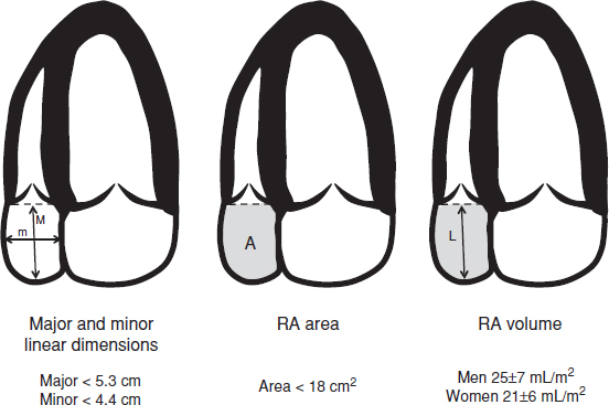
# Right Atrium
- 研究不如其他腔室充分
- Measurement of the right atrium is usually performed from the apical four-chamber or subcostal view.
右心房的测量通常从顶端四室或肋下视图进行。 - Linear dimensions can be determined, and the normal range of right atrial size has been reported.
线性距离是可以确定的,右房大小的正常范围已有报道。 - Planimetry of right atrial area should also be performed to more directly assess chamber area and volume.
还应进行右房面积测量,以更直接地评估房面积和容积。 - These measurements are performed at end-systole, prior to tricuspid valve opening.
这些测量是在三尖瓣打开之前的收缩末期进行的。 - This method is similar to that described earlier for the left atrium
这种方法与前面描述的左心房的方法相似 - The formula used to estimate right atrial volume from the four-chamber view is the single plane area-length method
从四腔心切面估计右心房体积的公式是单平面面积长度法

# Q&A
在某些病人,左室容积减小,用 tTVI 和 Simpson 法测得的左室 SV 不一致,应该 Simpson 法测量更准确一些吗?
对于 Simpson 法来说我们更关注这个方法测得的左室射血分数,也就是左室收缩的强度。对于每搏量来说,Simpson 法用的是减法,而 VTI 是通过区域流动的血流量和速度算得的,两个方法测出来的值可能不一样。但是 Simpson 方法是建立在室腔为特定形状的假设之上的,如果形状不符合这个假设时,就会产生一定的误差。而 VTI 的主要困难点是左室流出道内径的测量。两个方法各有优缺点。对于具体的某个病例,我们很难确定两种方法的好坏。如果患者 4C 或 2C 下心内膜的边界不是很清楚,Simpson 误差较大;同时患者左室流出道测量若比较精确,或者得到了比较好的 VTI,那么 VTI 的方法会更好一些。